 Movie
Movie Controller

[English] 日本語
 Yorodumi
- PDB-1odn: ISOPENICILLIN N SYNTHASE FROM ASPERGILLUS NIDULANS (OXYGEN-EXPOSE... -
+ Open data

- Basic information
| Entry | Database: PDB / ID: 1odn | ||||||
|---|---|---|---|---|---|---|---|
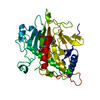
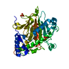
| Title | ISOPENICILLIN N SYNTHASE FROM ASPERGILLUS NIDULANS (OXYGEN-EXPOSED PRODUCT FROM ANAEROBIC AC-VINYLGLYCINE FE COMPLEX) | ||||||
 Components Components | ISOPENICILLIN N SYNTHASE | ||||||
 Keywords Keywords | OXIDOREDUCTASE / ANTIBIOTIC BIOSYNTHESIS / B-LACTAM ANTIBIOTIC / OXYGENASE / PENICILLIN BIOSYNTHESIS | ||||||
| Function / homology |  Function and homology information Function and homology informationisopenicillin-N synthase / isopenicillin-N synthase activity / penicillin biosynthetic process / L-ascorbic acid binding / iron ion binding / cytosol Similarity search - Function | ||||||
| Biological species |  | ||||||
| Method |  X-RAY DIFFRACTION / SYNCHROTRON / MOLECULAR REPLACEMENT / Resolution: 1.6 Å X-RAY DIFFRACTION / SYNCHROTRON / MOLECULAR REPLACEMENT / Resolution: 1.6 Å | ||||||
 Authors Authors | Elkins, J.M. / Rutledge, P.J. / Burzlaff, N.I. / Clifton, I.J. / Adlington, R.M. / Roach, P.L. / Baldwin, J.E. | ||||||
 Citation Citation | Journal: Org.Biomol.Chem. / Year: 2003 Title: Crystallographic Studies on the Reaction of Isopenicillin N Synthase with an Unsaturated Substrate Analogue Authors: Elkins, J.M. / Rutledge, P.J. / Burzlaff, N.I. / Clifton, I.J. / Adlington, R.M. / Roach, P.L. / Baldwin, J.E. #1: Journal: Nature / Year: 1999Title: The Reaction Cycle of Isopenicillin N Synthase Observed by X-Ray Diffraction Authors: Burzlaff, N.I. / Rutledge, P.J. / Clifton, I.J. / Hensgens, C.M.H. / Pickford, M. / Adlington, R.M. / Roach, P.L. / Baldwin, J.E. #2: Journal: Nature / Year: 1997Title: Structure of Isopenicillin N Synthase Complexed with Substrate and the Mechanism of Penicillin Formation Authors: Roach, P.L. / Clifton, I.J. / Hensgens, C.M. / Shibata, N. / Schofield, C.J. / Hajdu, J. / Baldwin, J.E. #3: Journal: Nature / Year: 1995Title: Crystal Structure of Isopenicillin N Synthase is the First from a New Structural Family of Enzymes Authors: Roach, P.L. / Clifton, I.J. / Fulop, V. / Harlos, K. / Barton, G.J. / Hajdu, J. / Andersson, I. / Schofield, C.J. / Baldwin, J.E. | ||||||
| History |
|
- Structure visualization
| Structure viewer | Molecule: MolmilJmol/JSmol |
|---|
- Downloads & links
-Download
| PDBx/mmCIF format | 1odn.cif.gz | 88.3 KB | Display | PDBx/mmCIF format |
|---|---|---|---|---|
| PDB format | pdb1odn.ent.gz | 66.2 KB | Display | PDB format |
| PDBx/mmJSON format | 1odn.json.gz | Tree view | PDBx/mmJSON format | |
| Others |  Other downloads Other downloads |
-Validation report
| Arichive directory | https://data.pdbj.org/pub/pdb/validation_reports/od/1odnftp://data.pdbj.org/pub/pdb/validation_reports/od/1odn | HTTPS FTP |
|---|
-Related structure data
| Related structure data |  1odmC  1qjeS C: citing same article ( S: Starting model for refinement |
|---|---|
| Similar structure data |


-Links
 PDBj
PDBj


- Assembly
| Deposited unit | 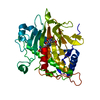
| ||||||||
|---|---|---|---|---|---|---|---|---|---|
| 1 |
| ||||||||
| Unit cell |
|
-Components
| #1: Protein | Mass: 37563.836 Da / Num. of mol.: 1 Source method: isolated from a genetically manipulated source Source: (gene. exp.) Plasmid: PJB703 / Production host:  |
|---|---|
| #2: Chemical | ChemComp-APV /   Mass: 361.371 Da / Num. of mol.: 1 / Source method: obtained synthetically / Formula: C13H19N3O7S Mass: 361.371 Da / Num. of mol.: 1 / Source method: obtained synthetically / Formula: C13H19N3O7S |
| #3: Chemical | ChemComp-SO4 /   Mass: 96.063 Da / Num. of mol.: 1 / Source method: obtained synthetically / Formula: SO4 Mass: 96.063 Da / Num. of mol.: 1 / Source method: obtained synthetically / Formula: SO4 |
| #4: Chemical | ChemComp-FE2 /   Mass: 55.845 Da / Num. of mol.: 1 / Source method: obtained synthetically / Formula: Fe Mass: 55.845 Da / Num. of mol.: 1 / Source method: obtained synthetically / Formula: Fe |
| #5: Water | ChemComp-HOH /  Mass: 18.015 Da / Num. of mol.: 395 / Source method: isolated from a natural source / Formula: H2O Mass: 18.015 Da / Num. of mol.: 395 / Source method: isolated from a natural source / Formula: H2O |
-Experimental details
-Experiment
| Experiment | Method: X-RAY DIFFRACTION / Number of used crystals: 1 |
|---|
- Sample preparation
| Crystal | Density Matthews: 2.26 Å3/Da / Density % sol: 40 % | |||||||||||||||
|---|---|---|---|---|---|---|---|---|---|---|---|---|---|---|---|---|
| Crystal grow | pH: 7.5 Details: 1.8M LITHIUM SULPHATE, 100MM TRIS/HCL (PH8.5), (5MM FERROUS SULPHATE, 70 MM ACVG, 50MG/ML IPNS), pH 7.50 | |||||||||||||||
| Crystal grow | *PLUS Temperature: 17 ℃ / pH: 8.5 / Method: vapor diffusion, hanging drop / Details: Roach, P.L., (1996) Eur. J. Biochem., 242, 736. | |||||||||||||||
| Components of the solutions | *PLUS
|
-Data collection
| Diffraction | Mean temperature: 100 K |
|---|---|
| Diffraction source | Source: SYNCHROTRON / Site: SRS  / Beamline: PX7.2 / Wavelength: 0.9312 / Beamline: PX7.2 / Wavelength: 0.9312 |
| Detector | Type: MARRESEARCH / Detector: IMAGE PLATE / Date: Apr 4, 1998 |
| Radiation | Protocol: SINGLE WAVELENGTH / Monochromatic (M) / Laue (L): M / Scattering type: x-ray |
| Radiation wavelength | Wavelength: 0.9312 Å / Relative weight: 1 |
| Reflection | Resolution: 1.6→41.17 Å / Num. obs: 42669 / % possible obs: 95.2 % / Redundancy: 2.4 % / Rmerge(I) obs: 0.074 / Net I/σ(I): 6.4 |
| Reflection shell | Resolution: 1.6→1.69 Å / Redundancy: 2.4 % / Rmerge(I) obs: 0.338 / Mean I/σ(I) obs: 1.9 / % possible all: 94.2 |
| Reflection | *PLUS Highest resolution: 1.6 Å / Lowest resolution: 41.17 Å / Redundancy: 2.4 % / Num. measured all: 101990 / Rmerge(I) obs: 0.074 |
| Reflection shell | *PLUS % possible obs: 94.2 % / Redundancy: 2.4 % / Num. unique obs: 6077 / Num. measured obs: 14387 / Rmerge(I) obs: 0.338 / Mean I/σ(I) obs: 1.9 |
- Processing
| Software |
| ||||||||||||||||||||||||||||||||||||||||||||||||||||||||||||||||||||||||||||||||||||||||||||||||||||||||||||||||||||||||||||||||||||||||||||||||||||||||||||||||||||||||||||||||||||||
|---|---|---|---|---|---|---|---|---|---|---|---|---|---|---|---|---|---|---|---|---|---|---|---|---|---|---|---|---|---|---|---|---|---|---|---|---|---|---|---|---|---|---|---|---|---|---|---|---|---|---|---|---|---|---|---|---|---|---|---|---|---|---|---|---|---|---|---|---|---|---|---|---|---|---|---|---|---|---|---|---|---|---|---|---|---|---|---|---|---|---|---|---|---|---|---|---|---|---|---|---|---|---|---|---|---|---|---|---|---|---|---|---|---|---|---|---|---|---|---|---|---|---|---|---|---|---|---|---|---|---|---|---|---|---|---|---|---|---|---|---|---|---|---|---|---|---|---|---|---|---|---|---|---|---|---|---|---|---|---|---|---|---|---|---|---|---|---|---|---|---|---|---|---|---|---|---|---|---|---|---|---|---|---|
| Refinement | Method to determine structure: MOLECULAR REPLACEMENT Starting model: PDB ENTRY 1QJE Resolution: 1.6→38 Å / Cor.coef. Fo:Fc: 0.97 / Cor.coef. Fo:Fc free: 0.959 / SU B: 1.359 / SU ML: 0.048 / TLS residual ADP flag: LIKELY RESIDUAL / Cross valid method: THROUGHOUT / ESU R: 0.081 / ESU R Free: 0.08 / Stereochemistry target values: MAXIMUM LIKELIHOOD / Details: HYDROGENS HAVE BEEN ADDED IN THE RIDING POSITIONS
| ||||||||||||||||||||||||||||||||||||||||||||||||||||||||||||||||||||||||||||||||||||||||||||||||||||||||||||||||||||||||||||||||||||||||||||||||||||||||||||||||||||||||||||||||||||||
| Solvent computation | Ion probe radii: 0.8 Å / Shrinkage radii: 0.8 Å / VDW probe radii: 1.4 Å / Solvent model: BABINET MODEL WITH MASK | ||||||||||||||||||||||||||||||||||||||||||||||||||||||||||||||||||||||||||||||||||||||||||||||||||||||||||||||||||||||||||||||||||||||||||||||||||||||||||||||||||||||||||||||||||||||
| Displacement parameters | Biso mean: 11.97 Å2
| ||||||||||||||||||||||||||||||||||||||||||||||||||||||||||||||||||||||||||||||||||||||||||||||||||||||||||||||||||||||||||||||||||||||||||||||||||||||||||||||||||||||||||||||||||||||
| Refinement step | Cycle: LAST / Resolution: 1.6→38 Å
| ||||||||||||||||||||||||||||||||||||||||||||||||||||||||||||||||||||||||||||||||||||||||||||||||||||||||||||||||||||||||||||||||||||||||||||||||||||||||||||||||||||||||||||||||||||||
| Refine LS restraints |
|