 Movie
Movie Controller

[English] 日本語
 Yorodumi
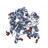
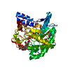
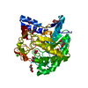
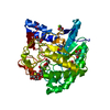
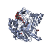
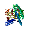
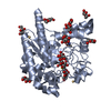
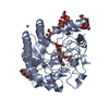
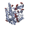
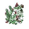
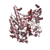
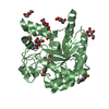
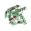
- PDB-1ocn: Mutant D416A of the CELLOBIOHYDROLASE CEL6A FROM HUMICOLA INSOLEN... -
+ Open data

- Basic information
| Entry | Database: PDB / ID: 1ocn | ||||||
|---|---|---|---|---|---|---|---|
| Title | Mutant D416A of the CELLOBIOHYDROLASE CEL6A FROM HUMICOLA INSOLENS in complex with a cellobio-derived isofagomine at 1.3 angstrom resolution | ||||||
 Components Components | CELLOBIOHYDROLASE II | ||||||
 Keywords Keywords | HYDROLASE / CELLULOSE DEGRADATION / CELLOBIOHYDROLASE / CELLULASE / GLYCOSIDE HYDROLASE FAMILY 6 | ||||||
| Function / homology |  Function and homology information Function and homology informationcellulose 1,4-beta-cellobiosidase (non-reducing end) / cellulose 1,4-beta-cellobiosidase activity / cellulose binding / cellulose catabolic process / extracellular region Similarity search - Function | ||||||
| Biological species |  HUMICOLA INSOLENS (fungus) HUMICOLA INSOLENS (fungus) | ||||||
| Method |  X-RAY DIFFRACTION / SYNCHROTRON / MOLECULAR REPLACEMENT / Resolution: 1.31 Å X-RAY DIFFRACTION / SYNCHROTRON / MOLECULAR REPLACEMENT / Resolution: 1.31 Å | ||||||
 Authors Authors | Varrot, A. / Macdonald, J. / Stick, R.V. / Pell, G. / Gilbert, H.J. / Davies, G.J. | ||||||
 Citation Citation | Journal: Chem.Commun.(Camb.) / Year: 2003 Title: Distortion of a Cellobio-Derived Isofagomine Highlights the Potential Conformational Itinerary of Inverting Beta-Glucosidases Authors: Varrot, A. / Macdonald, J. / Stick, R.V. / Pell, G. / Gilbert, H.J. / Davies, G.J. | ||||||
| History |
| ||||||
| Remark 700 | SHEET THE SHEET STRUCTURE OF THIS MOLECULE IS BIFURCATED. IN ORDER TO REPRESENT THIS FEATURE IN ... SHEET THE SHEET STRUCTURE OF THIS MOLECULE IS BIFURCATED. IN ORDER TO REPRESENT THIS FEATURE IN THE SHEET RECORDS BELOW, TWO SHEETS ARE DEFINED. |
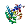
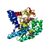
- Structure visualization
| Structure viewer | Molecule: MolmilJmol/JSmol |
|---|
- Downloads & links
-Download
| PDBx/mmCIF format | 1ocn.cif.gz | 176.9 KB | Display | PDBx/mmCIF format |
|---|---|---|---|---|
| PDB format | pdb1ocn.ent.gz | 138.7 KB | Display | PDB format |
| PDBx/mmJSON format | 1ocn.json.gz | Tree view | PDBx/mmJSON format | |
| Others |  Other downloads Other downloads |
-Validation report
| Arichive directory | https://data.pdbj.org/pub/pdb/validation_reports/oc/1ocnftp://data.pdbj.org/pub/pdb/validation_reports/oc/1ocn | HTTPS FTP |
|---|
-Related structure data
| Related structure data | 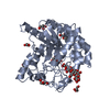 2bvwS S: Starting model for refinement |
|---|---|
| Similar structure data |


-Links
 PDBj
PDBj


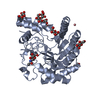
- Assembly
| Deposited unit | 
| ||||||||
|---|---|---|---|---|---|---|---|---|---|
| 1 |
| ||||||||
| Unit cell |
|
-Components
-Protein , 1 types, 1 molecules A
| #1: Protein | Mass: 39970.504 Da / Num. of mol.: 1 / Fragment: CATALYTIC CORE DOMAIN, RESIDUES 89-450 / Mutation: YES Source method: isolated from a genetically manipulated source Details: N-LINKED N-ACETYLGLUCOSAMINE ON RESIDUE ASN 141 / Source: (gene. exp.) HUMICOLA INSOLENS (fungus)Plasmid: UNDER CONTROL OF THE FUNGAL AMYLASE PROMOTER AND AMYLOGLUCOSIDASE TERMINATOR Production host: References: UniProt: Q9C1S9*PLUS, cellulose 1,4-beta-cellobiosidase (non-reducing end) |
|---|
-Sugars , 2 types, 3 molecules 

| #2: Sugar | ChemComp-NAG /  Type: D-saccharide, beta linking / Mass: 221.208 Da / Num. of mol.: 1 Type: D-saccharide, beta linking / Mass: 221.208 Da / Num. of mol.: 1Source method: isolated from a genetically manipulated source Formula: C8H15NO6 |
|---|---|
| #4: Sugar |  Type: D-saccharide, beta linking / Mass: 180.156 Da / Num. of mol.: 2 Type: D-saccharide, beta linking / Mass: 180.156 Da / Num. of mol.: 2Source method: isolated from a genetically manipulated source Formula: C6H12O6 |
-Non-polymers , 3 types, 525 molecules 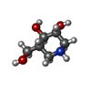


| #3: Chemical |  Mass: 147.172 Da / Num. of mol.: 2 / Source method: obtained synthetically / Formula: C6H13NO3 Mass: 147.172 Da / Num. of mol.: 2 / Source method: obtained synthetically / Formula: C6H13NO3#5: Chemical |  Mass: 40.078 Da / Num. of mol.: 2 / Source method: obtained synthetically / Formula: Ca Mass: 40.078 Da / Num. of mol.: 2 / Source method: obtained synthetically / Formula: Ca#6: Water | ChemComp-HOH / | Mass: 18.015 Da / Num. of mol.: 521 / Source method: isolated from a natural source / Formula: H2O |
|---|
-Details
| Compound details | ENGINEERED| Has protein modification | Y | Sequence details | THIS MUTANT HAS BEEN PRODUCED BY SITE-DIRECTED MUTAGENESIS. THE CLONING WAS PERFORMED SUCH HAS ONLY ...THIS MUTANT HAS BEEN PRODUCED BY SITE-DIRECTED MUTAGENESI | |
|---|
-Experimental details
-Experiment
| Experiment | Method: X-RAY DIFFRACTION / Number of used crystals: 1 |
|---|
- Sample preparation
| Crystal | Density Matthews: 2.01 Å3/Da / Density % sol: 42 % / Description: MOLECULE A |
|---|---|
| Crystal grow | pH: 7.5 Details: PROTEIN WAS CONCENTRATED TO 10MG/ML IN WATER.CRYSTALLISATION IN 200MM CALCIUM ACETATE, 100MM HEPES PH 7.5 AND 21% POLYETHYLENE GLYCOL 5KMME.20 % GLYCEROL WAS ADDED FOR CRYOPROTECTION |
-Data collection
| Diffraction | Mean temperature: 100 K |
|---|---|
| Diffraction source | Source: SYNCHROTRON / Site: ESRF  / Beamline: ID14-1 / Wavelength: 0.934 / Beamline: ID14-1 / Wavelength: 0.934 |
| Detector | Type: ADSC CCD / Detector: CCD / Date: Jun 15, 2002 / Details: TORROIDAL MIRROR |
| Radiation | Monochromator: DIAMOND (111), GE(220) / Protocol: SINGLE WAVELENGTH / Monochromatic (M) / Laue (L): M / Scattering type: x-ray |
| Radiation wavelength | Wavelength: 0.934 Å / Relative weight: 1 |
| Reflection | Resolution: 1.31→15 Å / Num. obs: 77018 / % possible obs: 91.7 % / Observed criterion σ(I): 0 / Redundancy: 6.2 % / Rmerge(I) obs: 0.06 / Net I/σ(I): 17.9 |
| Reflection shell | Resolution: 1.31→1.33 Å / Redundancy: 5.6 % / Rmerge(I) obs: 0.275 / Mean I/σ(I) obs: 7.6 / % possible all: 100 |
- Processing
| Software |
| ||||||||||||||||||||||||||||||||||||||||||||||||||||||||||||||||||||||||||||||||||||||||||||||||||||||||||||||||||||||||||||||||||||||||||||||||||||||||||||||||||||||||||||||||||||||
|---|---|---|---|---|---|---|---|---|---|---|---|---|---|---|---|---|---|---|---|---|---|---|---|---|---|---|---|---|---|---|---|---|---|---|---|---|---|---|---|---|---|---|---|---|---|---|---|---|---|---|---|---|---|---|---|---|---|---|---|---|---|---|---|---|---|---|---|---|---|---|---|---|---|---|---|---|---|---|---|---|---|---|---|---|---|---|---|---|---|---|---|---|---|---|---|---|---|---|---|---|---|---|---|---|---|---|---|---|---|---|---|---|---|---|---|---|---|---|---|---|---|---|---|---|---|---|---|---|---|---|---|---|---|---|---|---|---|---|---|---|---|---|---|---|---|---|---|---|---|---|---|---|---|---|---|---|---|---|---|---|---|---|---|---|---|---|---|---|---|---|---|---|---|---|---|---|---|---|---|---|---|---|---|
| Refinement | Method to determine structure: MOLECULAR REPLACEMENT Starting model: PDB ENTRY 2BVW Resolution: 1.31→15 Å / Cor.coef. Fo:Fc: 0.979 / Cor.coef. Fo:Fc free: 0.973 / SU B: 0.638 / SU ML: 0.028 / Cross valid method: THROUGHOUT / ESU R: 0.056 / ESU R Free: 0.051 / Stereochemistry target values: MAXIMUM LIKELIHOOD / Details: HYDROGENS HAVE BEEN ADDED IN THE RIDING POSITIONS
| ||||||||||||||||||||||||||||||||||||||||||||||||||||||||||||||||||||||||||||||||||||||||||||||||||||||||||||||||||||||||||||||||||||||||||||||||||||||||||||||||||||||||||||||||||||||
| Solvent computation | Ion probe radii: 0.8 Å / Shrinkage radii: 0.8 Å / VDW probe radii: 1.4 Å / Solvent model: BABINET MODEL WITH MASK | ||||||||||||||||||||||||||||||||||||||||||||||||||||||||||||||||||||||||||||||||||||||||||||||||||||||||||||||||||||||||||||||||||||||||||||||||||||||||||||||||||||||||||||||||||||||
| Displacement parameters | Biso mean: 12.29 Å2
| ||||||||||||||||||||||||||||||||||||||||||||||||||||||||||||||||||||||||||||||||||||||||||||||||||||||||||||||||||||||||||||||||||||||||||||||||||||||||||||||||||||||||||||||||||||||
| Refinement step | Cycle: LAST / Resolution: 1.31→15 Å
| ||||||||||||||||||||||||||||||||||||||||||||||||||||||||||||||||||||||||||||||||||||||||||||||||||||||||||||||||||||||||||||||||||||||||||||||||||||||||||||||||||||||||||||||||||||||
| Refine LS restraints |
|