 Movie
Movie Controller

[English] 日本語
 Yorodumi
- PDB-1obf: The crystal structure of Glyceraldehyde 3-phosphate Dehydrogenase... -
+ Open data

- Basic information
| Entry | Database: PDB / ID: 1obf | ||||||
|---|---|---|---|---|---|---|---|
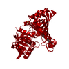
| Title | The crystal structure of Glyceraldehyde 3-phosphate Dehydrogenase from Alcaligenes xylosoxidans at 1.7A resolution. | ||||||
 Components Components | GLYCERALDEHYDE 3-PHOSPHATE DEHYDROGENASE | ||||||
 Keywords Keywords | GLYCOLYTIC PATHWAY / OXIDOREDUCTASE / FREE-NAD GAPDH | ||||||
| Function / homology |  Function and homology information Function and homology informationOxidoreductases; Acting on the aldehyde or oxo group of donors; With NAD+ or NADP+ as acceptor / oxidoreductase activity, acting on the aldehyde or oxo group of donors, NAD or NADP as acceptor / glucose metabolic process / NAD binding / NADP binding Similarity search - Function | ||||||
| Biological species |  ACHROMOBACTER XYLOSOXIDANS (bacteria) ACHROMOBACTER XYLOSOXIDANS (bacteria) | ||||||
| Method |  X-RAY DIFFRACTION / SYNCHROTRON / MOLECULAR REPLACEMENT / Resolution: 1.7 Å X-RAY DIFFRACTION / SYNCHROTRON / MOLECULAR REPLACEMENT / Resolution: 1.7 Å | ||||||
 Authors Authors | Antonyuk, S.V. / Eady, R.R. / Strange, R.W. / Hasnain, S.S. | ||||||
 Citation Citation | Journal: Acta Crystallogr.,Sect.D / Year: 2003 Title: The Structure of Glyceraldehyde 3-Phosphate Dehydrogenase from Alcaligenes Xylosoxidans at 1.7 A Resolution Authors: Antonyuk, S.V. / Eady, R.R. / Strange, R.W. / Hasnain, S.S. | ||||||
| History |
| ||||||
| Remark 700 | SHEET THE SHEET STRUCTURE OF THIS MOLECULE IS BIFURCATED. IN ORDER TO REPRESENT THIS FEATURE IN ... SHEET THE SHEET STRUCTURE OF THIS MOLECULE IS BIFURCATED. IN ORDER TO REPRESENT THIS FEATURE IN THE SHEET RECORDS BELOW, TWO SHEETS ARE DEFINED. |
- Structure visualization
| Structure viewer | Molecule: MolmilJmol/JSmol |
|---|
- Downloads & links
-Download
| PDBx/mmCIF format | 1obf.cif.gz | 145.6 KB | Display | PDBx/mmCIF format |
|---|---|---|---|---|
| PDB format | pdb1obf.ent.gz | 114.4 KB | Display | PDB format |
| PDBx/mmJSON format | 1obf.json.gz | Tree view | PDBx/mmJSON format | |
| Others |  Other downloads Other downloads |
-Validation report
| Arichive directory | https://data.pdbj.org/pub/pdb/validation_reports/ob/1obfftp://data.pdbj.org/pub/pdb/validation_reports/ob/1obf | HTTPS FTP |
|---|
-Related structure data
| Related structure data |  1gd1S S: Starting model for refinement |
|---|---|
| Similar structure data |


-Links
 PDBj
PDBj
- Assembly
| Deposited unit | 
| ||||||||
|---|---|---|---|---|---|---|---|---|---|
| 1 | 
| ||||||||
| Unit cell |
| ||||||||
| Components on special symmetry positions |
| ||||||||
| Noncrystallographic symmetry (NCS) | NCS oper: (Code: given Matrix: (0.989, 0.003, -0.151), Vector: |
-Components
| #1: Protein | Mass: 35828.441 Da / Num. of mol.: 2 / Source method: isolated from a natural source / Source: (natural) ACHROMOBACTER XYLOSOXIDANS (bacteria)References: UniProt: P83696*PLUS, glyceraldehyde-3-phosphate dehydrogenase (phosphorylating) #2: Chemical |   Mass: 96.063 Da / Num. of mol.: 3 / Source method: obtained synthetically / Formula: SO4 Mass: 96.063 Da / Num. of mol.: 3 / Source method: obtained synthetically / Formula: SO4#3: Chemical |   Mass: 39.098 Da / Num. of mol.: 2 / Source method: obtained synthetically / Formula: K Mass: 39.098 Da / Num. of mol.: 2 / Source method: obtained synthetically / Formula: K#4: Chemical | ChemComp-PG4 / |   Mass: 194.226 Da / Num. of mol.: 1 / Source method: obtained synthetically / Formula: C8H18O5 / Comment: precipitant*YM Mass: 194.226 Da / Num. of mol.: 1 / Source method: obtained synthetically / Formula: C8H18O5 / Comment: precipitant*YM#5: Water | ChemComp-HOH / |  Mass: 18.015 Da / Num. of mol.: 501 / Source method: isolated from a natural source / Formula: H2O Mass: 18.015 Da / Num. of mol.: 501 / Source method: isolated from a natural source / Formula: H2OHas protein modification | Y | |
|---|
-Experimental details
-Experiment
| Experiment | Method: X-RAY DIFFRACTION / Number of used crystals: 1 |
|---|
- Sample preparation
| Crystal | Density Matthews: 3.3 Å3/Da / Density % sol: 59 % | ||||||||||||||||||||||||||||||||||||||||||||||||
|---|---|---|---|---|---|---|---|---|---|---|---|---|---|---|---|---|---|---|---|---|---|---|---|---|---|---|---|---|---|---|---|---|---|---|---|---|---|---|---|---|---|---|---|---|---|---|---|---|---|
| Crystal grow | pH: 6.5 Details: PROTEIN 5MG/ML, 12 % PEG550, 100 MM POTASSIUM THIOCYANATE, 50 MM MES BUFFER (PH 6.5) | ||||||||||||||||||||||||||||||||||||||||||||||||
| Crystal grow | *PLUS pH: 6.5 / Method: vapor diffusion, hanging drop | ||||||||||||||||||||||||||||||||||||||||||||||||
| Components of the solutions | *PLUS
|
-Data collection
| Diffraction | Mean temperature: 100 K |
|---|---|
| Diffraction source | Source: SYNCHROTRON / Site: SRS  / Beamline: PX9.6 / Wavelength: 0.87 / Beamline: PX9.6 / Wavelength: 0.87 |
| Detector | Type: ADSC CCD / Detector: CCD / Date: May 15, 2001 / Details: MIRRORS |
| Radiation | Monochromator: NI FILTER / Protocol: SINGLE WAVELENGTH / Monochromatic (M) / Laue (L): M / Scattering type: x-ray |
| Radiation wavelength | Wavelength: 0.87 Å / Relative weight: 1 |
| Reflection | Resolution: 1.7→50 Å / Num. obs: 102122 / % possible obs: 97.6 % / Redundancy: 6.7 % / Biso Wilson estimate: 22.7 Å2 / Rmerge(I) obs: 0.099 / Net I/σ(I): 22 |
| Reflection shell | Resolution: 1.7→1.76 Å / Redundancy: 6 % / Rmerge(I) obs: 0.45 / Mean I/σ(I) obs: 2.2 / % possible all: 90.7 |
| Reflection | *PLUS Highest resolution: 1.7 Å / Lowest resolution: 50 Å / Num. obs: 104690 / Redundancy: 6 % / Rmerge(I) obs: 0.099 |
| Reflection shell | *PLUS Highest resolution: 1.7 Å / % possible obs: 90.7 % / Rmerge(I) obs: 0.35 / Mean I/σ(I) obs: 2.2 |
- Processing
| Software |
| ||||||||||||||||||||
|---|---|---|---|---|---|---|---|---|---|---|---|---|---|---|---|---|---|---|---|---|---|
| Refinement | Method to determine structure: MOLECULAR REPLACEMENT Starting model: PDB ENTRY 1GD1 Resolution: 1.7→50 Å / SU B: 1.871 / SU ML: 0.058 / Cross valid method: THROUGHOUT / ESU R: 0.079 / ESU R Free: 0.078 / Details: HYDROGENS HAVE BEEN ADDED IN THE RIDING POSITIONS
| ||||||||||||||||||||
| Displacement parameters | Biso mean: 23.299 Å2
| ||||||||||||||||||||
| Refinement step | Cycle: LAST / Resolution: 1.7→50 Å
| ||||||||||||||||||||
| Refinement | *PLUS Lowest resolution: 50 Å / Rfactor Rfree: 0.189 / Rfactor Rwork: 0.168 | ||||||||||||||||||||
| Solvent computation | *PLUS | ||||||||||||||||||||
| Displacement parameters | *PLUS | ||||||||||||||||||||
| Refine LS restraints | *PLUS
|
