Mass: 96.063 Da / Num. of mol.: 2 / Source method: obtained synthetically / Formula: SO4
Compound details
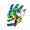
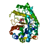
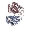
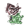
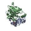
CHEMOTACTIC FOR MONOCYTES AND T LYMPHOCYTES. BINDS TO CXCR3. INDUCED BY INTERFERON GAMMA. A DIVERSE ...CHEMOTACTIC FOR MONOCYTES AND T LYMPHOCYTES. BINDS TO CXCR3. INDUCED BY INTERFERON GAMMA. A DIVERSE POPULATION OF CELL TYPES RAPIDLY INCREASES TRANSCRIPTION OF MRNA ENCODING THIS PROTEIN. THIS SUGGESTS THAT GAMMA-INDUCED PROTEIN MAY BE A KEY MEDIATOR OF THE INTERFERON GAMMA RESPONSE.
Has protein modification
Y
Sequence details
THE SEQUENCE CONFLICT INDICATED IN THE SEQADV RECORDS ARISES FROM A DIFFERENCE IN THE PRIMARY ...THE SEQUENCE CONFLICT INDICATED IN THE SEQADV RECORDS ARISES FROM A DIFFERENCE IN THE PRIMARY SEQUENCE IN THE SWISS-PROT DATABASE REFERENCE P02778 AT POSITION 93. THE SEQUENCE GIVEN HERE FOLLOWS THE SEQUENCE DESCRIBED IN REFERENCE: LUSTER ET AL., NATURE, 315:672 (1985).
-
Experimental details
-
Experiment
Experiment
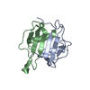
Method: X-RAY DIFFRACTION / Number of used crystals: 1
-
Sample preparation
Crystal
Density Matthews: 2.77 Å3/Da / Density % sol: 55.4 %
In the structure databanks used in Yorodumi, some data are registered as the other names, "COVID-19 virus" and "2019-nCoV". Here are the details of the virus and the list of structure data.
Jan 31, 2019. EMDB accession codes are about to change! (news from PDBe EMDB page)
EMDB accession codes are about to change! (news from PDBe EMDB page)
The allocation of 4 digits for EMDB accession codes will soon come to an end. Whilst these codes will remain in use, new EMDB accession codes will include an additional digit and will expand incrementally as the available range of codes is exhausted. The current 4-digit format prefixed with “EMD-” (i.e. EMD-XXXX) will advance to a 5-digit format (i.e. EMD-XXXXX), and so on. It is currently estimated that the 4-digit codes will be depleted around Spring 2019, at which point the 5-digit format will come into force.
The EM Navigator/Yorodumi systems omit the EMD- prefix.
Related info.:Q: What is EMD? / ID/Accession-code notation in Yorodumi/EM Navigator
Yorodumi is a browser for structure data from EMDB, PDB, SASBDB, etc.
This page is also the successor to EM Navigator detail page, and also detail information page/front-end page for Omokage search.
The word "yorodu" (or yorozu) is an old Japanese word meaning "ten thousand". "mi" (miru) is to see.
Related info.:EMDB / PDB / SASBDB / Comparison of 3 databanks / Yorodumi Search / Aug 31, 2016. New EM Navigator & Yorodumi / Yorodumi Papers / Jmol/JSmol / Function and homology information / Changes in new EM Navigator and Yorodumi
 Movie
Movie Controller


 Open data
 Basic information
 Components
 Keywords
 Function and homology information
 HOMO SAPIENS (human)
 X-RAY DIFFRACTION /
  Authors
 Citation
 Structure visualization
 Downloads & links
 Other downloads
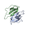


 PDBj
PDBj

 Assembly

 Mass: 96.063 Da / Num. of mol.: 2 / Source method: obtained synthetically / Formula: SO4
 Sample preparation
 / Beamline: X11 / Wavelength: 0.9057
  Processing