[English] 日本語
Yorodumi
- PDB-1o6a: CRYSTAL STRUCTURE OF A C-TERMINAL FRAGMENT OF THE PUTATIVE FLAGEL... -

+
Open data


ID or keywords:

Loading...

-
Basic information

Entry
Database: PDB / ID: 1o6a
Title
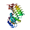
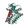
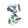
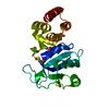
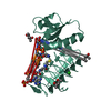
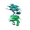
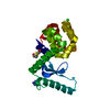
CRYSTAL STRUCTURE OF A C-TERMINAL FRAGMENT OF THE PUTATIVE FLAGELLAR MOTOR SWITCH PROTEIN FLIN (TM0680) FROM THERMOTOGA MARITIMA AT 1.85 A RESOLUTION
Componentsputative flagellar motor switch protein FliN
KeywordsMOTOR PROTEIN / PUTATIVE FLAGELLAR MOTOR SWITCH PROTEIN FLIN / C- TERMINAL PROTEOLYTIC FRAGMENT / STRUCTURAL GENOMICS / JOINT CENTER FOR STRUCTURAL GENOMICS / JCSG / PROTEIN STRUCTURE INITIATIVE / PSI
Function / homologySurface presentation of antigens (SPOA) / SpoA-like / Roll / Mainly Beta / :
Function and homology information
Biological speciesThermotoga maritima (bacteria)
MethodX-RAY DIFFRACTION / SYNCHROTRON / MAD / Resolution: 1.85 Å
AuthorsJoint Center for Structural Genomics (JCSG)
CitationJournal: To be published
Title: Crystal structure of the C-terminal fragment of the putative flagellar motor switch protein FliN (TM0680a) from Thermotoga maritima at 1.85 A resolution
Authors: Joint Center for Structural Genomics (JCSG)
History
DepositionOct 24, 2003Deposition site: RCSB / Processing site: RCSB
Revision 1.0Nov 11, 2003Provider: repository / Type: Initial release
Revision 1.1Apr 26, 2008Group: Version format compliance
Revision 1.2Jul 13, 2011Group: Advisory / Version format compliance
Revision 1.3Jul 18, 2018Group: Data collection / Database references / Category: pdbx_database_related
Revision 1.4Dec 27, 2023Group: Data collection / Database references ...Data collection / Database references / Derived calculations / Refinement description
Category: chem_comp_atom / chem_comp_bond ...chem_comp_atom / chem_comp_bond / database_2 / struct_conn / struct_ncs_dom_lim / struct_ref_seq_dif
Item: _database_2.pdbx_DOI / _database_2.pdbx_database_accession ..._database_2.pdbx_DOI / _database_2.pdbx_database_accession / _struct_conn.pdbx_leaving_atom_flag / _struct_ncs_dom_lim.beg_auth_comp_id / _struct_ncs_dom_lim.end_auth_comp_id / _struct_ref_seq_dif.details
Remark 999SEQUENCE IN BOTH CHAINS THE DENSITY AND REFINEMENT STATISTICS FOR THE WELL ORDERED REGION AROUND ...SEQUENCE IN BOTH CHAINS THE DENSITY AND REFINEMENT STATISTICS FOR THE WELL ORDERED REGION AROUND RESIDUE 100 SUGGESTS THAT RESIDUE 100 IS A HISTIDINE. THIS PROTEIN WAS EXPRESSED WITH A 201 RESIDUE N-TERMINAL FUSION COMPRISING A 12 AMINO ACID PURIFICATION TAG, TM0680 (GB 15644623 / NP_228488) AND A 27 RESIDUE LINKER, AND TM0680A. IT CAN BE REPRESENTED AS [HIS6TAG]-[THYOREDOXIN LEADER]- [FUSION PROTEIN, FLIY = TM0680]-[LINKER]-[TM0680A]. THE TAG, TM0680, THE 27 RESIDUE LINKER, AND THE FIRST 58 RESIDUES OF TM0680A WERE CLEAVED, LEAVING RESIDUES 59-154 OF TM0680A IN THE CRYSTAL.

-
Structure visualization

Structure viewerMolecule:
MolmilJmol/JSmol

Downloads & links

-
Assembly

Deposited unit
A: putative flagellar motor switch protein FliN
B: putative flagellar motor switch protein FliN


Theoretical massNumber of molelcules
Total (without water)21,8492
Polymers21,8492
Non-polymers00
Water2,702150
1


  • Idetical with deposited unit
  • defined by author&software
TypeNameSymmetry operationNumber
identity operation1_555x,y,z1
Buried area5300 Å2
ΔGint-41 kcal/mol
Surface area9380 Å2
MethodPISA
Unit cell
Length a, b, c (Å)61.481, 61.481, 113.148
Angle α, β, γ (deg.)90.00, 90.00, 90.00
Int Tables number92
Space group name H-MP41212
Noncrystallographic symmetry (NCS)NCS domain:
IDEns-IDDetails
11A
21B

NCS domain segments:

Component-ID: 1 / Ens-ID: 1 / Beg auth comp-ID: LEU / Beg label comp-ID: LEU / End auth comp-ID: GLU / End label comp-ID: GLU / Refine code: 5 / Auth seq-ID: 74 - 154 / Label seq-ID: 16 - 96

Dom-IDAuth asym-IDLabel asym-ID
1AA
2BB

-
Components

#1: Protein putative flagellar motor switch protein FliN


Mass: 10924.471 Da / Num. of mol.: 2 / Fragment: C-terminal fragment (residues 59-154)
Source method: isolated from a genetically manipulated source
Details: fusion protein, see remark 999 / Source: (gene. exp.) Thermotoga maritima (bacteria) / Gene: TM0680a / Production host: Escherichia coli (E. coli) / References: GenBank: 15644624
#2: Water ChemComp-HOH / water


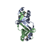
Mass: 18.015 Da / Num. of mol.: 150 / Source method: isolated from a natural source / Formula: H2O

-
Experimental details

-
Experiment

ExperimentMethod: X-RAY DIFFRACTION / Number of used crystals: 1

-
Sample preparation

CrystalDensity Matthews: 2.5 Å3/Da / Density % sol: 50.33 %
Crystal growTemperature: 273 K / Method: vapor diffusion, sitting drop, nanodrop / pH: 9
Details: 65% (+/-)-2-Methyl-2,4-Pentanediol, 0.1 M Bicine pH 9.0 , VAPOR DIFFUSION,SITTING DROP,NANODROP, temperature 273K

-
Data collection

DiffractionMean temperature: 100 K
Diffraction sourceSource: SYNCHROTRON / Site: ALS / Beamline: 8.3.1 / Wavelength: 1.019943, 0.979764, 0.979570
DetectorType: ADSC / Detector: CCD / Date: Jun 23, 2003
RadiationMonochromator: Double Crystal Si(111) / Protocol: MAD / Monochromatic (M) / Laue (L): M / Scattering type: x-ray
Radiation wavelength
IDWavelength (Å)Relative weight
11.0199431
20.9797641
30.979571
ReflectionResolution: 1.85→56.57 Å / Num. all: 19259 / Num. obs: 19259 / % possible obs: 99.9 % / Redundancy: 6.7 % / Biso Wilson estimate: 32.69 Å2 / Rsym value: 0.079 / Net I/σ(I): 16.4
Reflection shellResolution: 1.85→1.9 Å / Redundancy: 6.1 % / Mean I/σ(I) obs: 2.8 / Num. unique all: 1385 / Rsym value: 0.548 / % possible all: 100

-
Processing

Software
NameVersionClassification
MOSFLMdata reduction
SCALA4.2)data scaling
SOLVEphasing
RESOLVEmodel building
REFMAC5.1.24refinement
CCP4(SCALA)data scaling
RESOLVEphasing
RefinementMethod to determine structure: MAD / Resolution: 1.85→41.63 Å / Cor.coef. Fo:Fc: 0.961 / Cor.coef. Fo:Fc free: 0.947 / SU B: 2.454 / SU ML: 0.072 / TLS residual ADP flag: LIKELY RESIDUAL / Isotropic thermal model: ISOTROPIC / Cross valid method: THROUGHOUT / ESU R: 0.115 / ESU R Free: 0.116
Stereochemistry target values: MAXIMUM LIKELIHOOD WITH PHASES
Details: HYDROGENS HAVE BEEN ADDED IN THE RIDING POSITIONS.
RfactorNum. reflection% reflectionSelection details
Rfree0.21199 975 5.1 %RANDOM
Rwork0.17102 ---
obs0.17301 18226 99.85 %-
Solvent computationIon probe radii: 0.8 Å / Shrinkage radii: 0.8 Å / VDW probe radii: 1.4 Å / Solvent model: BABINET MODEL WITH MASK
Displacement parametersBiso mean: 18.923 Å2
Baniso -1Baniso -2Baniso -3
1-1.18 Å20 Å20 Å2
2--1.18 Å20 Å2
3----2.35 Å2
Refinement stepCycle: LAST / Resolution: 1.85→41.63 Å
ProteinNucleic acidLigandSolventTotal
Num. atoms1322 0 0 150 1472
Refine LS restraints
Refine-IDTypeDev idealDev ideal targetNumber
X-RAY DIFFRACTIONr_bond_refined_d0.0190.0221398
X-RAY DIFFRACTIONr_bond_other_d0.0020.021457
X-RAY DIFFRACTIONr_angle_refined_deg1.552.031903
X-RAY DIFFRACTIONr_angle_other_deg0.81533394
X-RAY DIFFRACTIONr_dihedral_angle_1_deg5.495186
X-RAY DIFFRACTIONr_chiral_restr0.0810.2251
X-RAY DIFFRACTIONr_gen_planes_refined0.0060.021485
X-RAY DIFFRACTIONr_gen_planes_other0.0020.02232
X-RAY DIFFRACTIONr_nbd_refined0.1930.2226
X-RAY DIFFRACTIONr_nbd_other0.2450.21774
X-RAY DIFFRACTIONr_nbtor_other0.0860.2974
X-RAY DIFFRACTIONr_xyhbond_nbd_refined0.2110.2109
X-RAY DIFFRACTIONr_symmetry_vdw_refined0.1510.27
X-RAY DIFFRACTIONr_symmetry_vdw_other0.4060.252
X-RAY DIFFRACTIONr_symmetry_hbond_refined0.2180.235
X-RAY DIFFRACTIONr_mcbond_it1.9243865
X-RAY DIFFRACTIONr_mcangle_it3.34951438
X-RAY DIFFRACTIONr_scbond_it6.098533
X-RAY DIFFRACTIONr_scangle_it8.65511455
Refine LS restraints NCS

Dom-ID: 1 / Auth asym-ID: A / Ens-ID: 1 / Refine-ID: X-RAY DIFFRACTION

NumberTypeRms dev position (Å)Weight position
474medium positional0.210.5
820loose positional0.535
474medium thermal0.982
820loose thermal1.8810
LS refinement shellResolution: 1.85→1.898 Å / Total num. of bins used: 20
RfactorNum. reflection% reflection
Rfree0.247 66 4.78 %
Rwork0.227 1316 -
Refinement TLS params.

Method: refined / Refine-ID: X-RAY DIFFRACTION

IDL112)L122)L132)L222)L232)L332)S11 (Å °)S12 (Å °)S13 (Å °)S21 (Å °)S22 (Å °)S23 (Å °)S31 (Å °)S32 (Å °)S33 (Å °)T112)T122)T132)T222)T232)T332)Origin x (Å)Origin y (Å)Origin z (Å)
11.16470.10220.16551.04121.00081.9129-0.00440.05690.203-0.19490.0697-0.0258-0.39770.1364-0.06530.063-0.01030.01120.0458-0.00380.084813.48831.00246.835
20.75310.1489-0.15040.76560.06870.51120.07480.10530.06740.01410.00810.1273-0.039-0.1677-0.0830.0210.0285-0.00930.1020.02440.078-9.40921.83740.338
32.01640.1386-0.50691.5747-0.01121.80670.0921-0.00280.17510.0797-0.05990.1096-0.2588-0.1827-0.03220.00670.01960.00040.0582-0.0040.0547-9.74724.0446.429
40.5654-0.09490.34571.02930.57030.95810.0931-0.0040.04380.01190.0497-0.1255-0.13270.2347-0.14280.0635-0.0224-0.00250.0944-0.03880.099312.89627.27352.532
Refinement TLS group

Refine-ID: X-RAY DIFFRACTION / Selection: ALL

IDRefine TLS-IDAuth asym-IDLabel asym-IDAuth seq-IDLabel seq-ID
11AA68 - 8410 - 26
21AA112 - 15454 - 96
32AA85 - 11127 - 53
43BB74 - 8416 - 26
53BB112 - 15454 - 96
64BB85 - 11127 - 53

+
About Yorodumi

-
News

-
Feb 9, 2022. New format data for meta-information of EMDB entries

New format data for meta-information of EMDB entries

  • Version 3 of the EMDB header file is now the official format.
  • The previous official version 1.9 will be removed from the archive.

Related info.:EMDB header

External links:wwPDB to switch to version 3 of the EMDB data model

-
Aug 12, 2020. Covid-19 info

Covid-19 info

URL: https://pdbjlc1.pdbj.org/emnavi/covid19.php

New page: Covid-19 featured information page in EM Navigator.

Related info.:Covid-19 info / Mar 5, 2020. Novel coronavirus structure data

+
Mar 5, 2020. Novel coronavirus structure data

Novel coronavirus structure data

Related info.:Yorodumi Speices / Aug 12, 2020. Covid-19 info

External links:COVID-19 featured content - PDBj / Molecule of the Month (242):Coronavirus Proteases

+
Jan 31, 2019. EMDB accession codes are about to change! (news from PDBe EMDB page)

EMDB accession codes are about to change! (news from PDBe EMDB page)

  • The allocation of 4 digits for EMDB accession codes will soon come to an end. Whilst these codes will remain in use, new EMDB accession codes will include an additional digit and will expand incrementally as the available range of codes is exhausted. The current 4-digit format prefixed with “EMD-” (i.e. EMD-XXXX) will advance to a 5-digit format (i.e. EMD-XXXXX), and so on. It is currently estimated that the 4-digit codes will be depleted around Spring 2019, at which point the 5-digit format will come into force.
  • The EM Navigator/Yorodumi systems omit the EMD- prefix.

Related info.:Q: What is EMD? / ID/Accession-code notation in Yorodumi/EM Navigator

External links:EMDB Accession Codes are Changing Soon! / Contact to PDBj

+
Jul 12, 2017. Major update of PDB

Major update of PDB

  • wwPDB released updated PDB data conforming to the new PDBx/mmCIF dictionary.
  • This is a major update changing the version number from 4 to 5, and with Remediation, in which all the entries are updated.
  • In this update, many items about electron microscopy experimental information are reorganized (e.g. em_software).
  • Now, EM Navigator and Yorodumi are based on the updated data.

External links:wwPDB Remediation / Enriched Model Files Conforming to OneDep Data Standards Now Available in the PDB FTP Archive

-
Yorodumi

Thousand views of thousand structures

  • Yorodumi is a browser for structure data from EMDB, PDB, SASBDB, etc.
  • This page is also the successor to EM Navigator detail page, and also detail information page/front-end page for Omokage search.
  • The word "yorodu" (or yorozu) is an old Japanese word meaning "ten thousand". "mi" (miru) is to see.

Related info.:EMDB / PDB / SASBDB / Comparison of 3 databanks / Yorodumi Search / Aug 31, 2016. New EM Navigator & Yorodumi / Yorodumi Papers / Jmol/JSmol / Function and homology information / Changes in new EM Navigator and Yorodumi

Read more