 Movie
Movie Controller

[English] 日本語
 Yorodumi
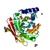
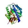
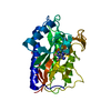
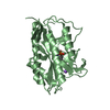
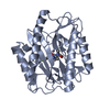
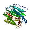
- PDB-1o0x: Crystal structure of Methionine aminopeptidase (TM1478) from Ther... -
+ Open data

- Basic information
| Entry | Database: PDB / ID: 1o0x | ||||||
|---|---|---|---|---|---|---|---|
| Title | Crystal structure of Methionine aminopeptidase (TM1478) from Thermotoga maritima at 1.90 A resolution | ||||||
 Components Components | Methionine aminopeptidase | ||||||
 Keywords Keywords | HYDROLASE / TM1478 / Methionine aminopeptidase / STRUCTURAL GENOMICS / JCSG / PSI / Protein Structure Initiative / Joint Center for Structural Genomics | ||||||
| Function / homology |  Function and homology information Function and homology informationmethionyl aminopeptidase / initiator methionyl aminopeptidase activity / metalloaminopeptidase activity / proteolysis / metal ion binding / cytosol Similarity search - Function | ||||||
| Biological species |   Thermotoga maritima (bacteria) Thermotoga maritima (bacteria) | ||||||
| Method |  X-RAY DIFFRACTION / SYNCHROTRON / MOLECULAR REPLACEMENT WITH PROGRAM AMORE / Resolution: 1.9 Å X-RAY DIFFRACTION / SYNCHROTRON / MOLECULAR REPLACEMENT WITH PROGRAM AMORE / Resolution: 1.9 Å | ||||||
 Authors Authors | Joint Center for Structural Genomics (JCSG) | ||||||
 Citation Citation | Journal: Proteins / Year: 2004 Title: Crystal structure of a methionine aminopeptidase (TM1478) from Thermotoga maritima at 1.9 A resolution. Authors: Spraggon, G. / Schwarzenbacher, R. / Kreusch, A. / McMullan, D. / Brinen, L.S. / Canaves, J.M. / Dai, X. / Deacon, A.M. / Elsliger, M.A. / Eshagi, S. / Floyd, R. / Godzik, A. / Grittini, C. ...Authors: Spraggon, G. / Schwarzenbacher, R. / Kreusch, A. / McMullan, D. / Brinen, L.S. / Canaves, J.M. / Dai, X. / Deacon, A.M. / Elsliger, M.A. / Eshagi, S. / Floyd, R. / Godzik, A. / Grittini, C. / Grzechnik, S.K. / Jaroszewski, L. / Karlak, C. / Klock, H.E. / Koesema, E. / Kovarik, J.S. / Kuhn, P. / McPhillips, T.M. / Miller, M.D. / Morse, A. / Moy, K. / Ouyang, J. / Page, R. / Quijano, K. / Rezezadeh, F. / Robb, A. / Sims, E. / Stevens, R.C. / van den Bedem, H. / Velasquez, J. / Vincent, J. / von Delft, F. / Wang, X. / West, B. / Wolf, G. / Xu, Q. / Hodgson, K.O. / Wooley, J. / Lesley, S.A. / Wilson, I.A. | ||||||
| History |
|
- Structure visualization
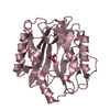
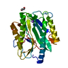
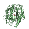
| Structure viewer | Molecule: MolmilJmol/JSmol |
|---|
- Downloads & links
-Download
| PDBx/mmCIF format | 1o0x.cif.gz | 67 KB | Display | PDBx/mmCIF format |
|---|---|---|---|---|
| PDB format | pdb1o0x.ent.gz | 47.9 KB | Display | PDB format |
| PDBx/mmJSON format | 1o0x.json.gz | Tree view | PDBx/mmJSON format | |
| Others |  Other downloads Other downloads |
-Validation report
| Arichive directory | https://data.pdbj.org/pub/pdb/validation_reports/o0/1o0xftp://data.pdbj.org/pub/pdb/validation_reports/o0/1o0x | HTTPS FTP |
|---|
-Related structure data
| Related structure data | 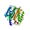 1matS S: Starting model for refinement |
|---|---|
| Similar structure data | |
| Other databases |

-Links
 PDBj
PDBj- Assembly
| Deposited unit | 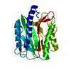
| ||||||||
|---|---|---|---|---|---|---|---|---|---|
| 1 |
| ||||||||
| Unit cell |
|
-Components
| #1: Protein | Mass: 28991.502 Da / Num. of mol.: 1 Source method: isolated from a genetically manipulated source Source: (gene. exp.) Thermotoga maritima (bacteria) / Gene: TM1478 / Production host: |
|---|---|
| #2: Water | ChemComp-HOH /  Mass: 18.015 Da / Num. of mol.: 195 / Source method: isolated from a natural source / Formula: H2O Mass: 18.015 Da / Num. of mol.: 195 / Source method: isolated from a natural source / Formula: H2O |
-Experimental details
-Experiment
| Experiment | Method: X-RAY DIFFRACTION / Number of used crystals: 1 |
|---|
- Sample preparation
| Crystal | Density Matthews: 2.47 Å3/Da / Density % sol: 50.14 % | |||||||||||||||||||||||||||||||||||||||||||||||||||||||||||||||
|---|---|---|---|---|---|---|---|---|---|---|---|---|---|---|---|---|---|---|---|---|---|---|---|---|---|---|---|---|---|---|---|---|---|---|---|---|---|---|---|---|---|---|---|---|---|---|---|---|---|---|---|---|---|---|---|---|---|---|---|---|---|---|---|---|
| Crystal grow | Temperature: 293 K / pH: 5.6 Details: 19% iso-propanol, 19% P4K, 0.095 M Na-citrate pH 5.6, 5% glycerol, VAPOR DIFFUSION,SITTING DROP,NANODROP, temperature 293K, pH 5.60 | |||||||||||||||||||||||||||||||||||||||||||||||||||||||||||||||
| Crystal grow | *PLUS pH: 7.9 / Method: vapor diffusion | |||||||||||||||||||||||||||||||||||||||||||||||||||||||||||||||
| Components of the solutions | *PLUS
|
-Data collection
| Diffraction | Mean temperature: 100 K |
|---|---|
| Diffraction source | Source: SYNCHROTRON / Site: ALS  / Beamline: 5.0.3 / Wavelength: 1 / Beamline: 5.0.3 / Wavelength: 1 |
| Detector | Type: ADSC QUANTUM 4 / Detector: CCD / Date: May 29, 2001 |
| Radiation | Monochromator: ASYMMETRICALLY CUT SI CRYSTAL, CYLINDRICALLY BENT Protocol: SINGLE WAVELENGTH / Monochromatic (M) / Laue (L): M / Scattering type: x-ray |
| Radiation wavelength | Wavelength: 1 Å / Relative weight: 1 |
| Reflection | Resolution: 1.9→50 Å / Num. obs: 21470 / % possible obs: 90.6 % / Biso Wilson estimate: 53 Å2 / Rsym value: 0.07 / Net I/σ(I): 16.53 |
| Reflection shell | Resolution: 1.9→1.97 Å / Mean I/σ(I) obs: 2.95 / % possible all: 90 |
| Reflection | *PLUS Highest resolution: 1.9 Å / Lowest resolution: 50 Å / Num. measured all: 102128 / Rmerge(I) obs: 0.07 |
| Reflection shell | *PLUS % possible obs: 90 % / Rmerge(I) obs: 0.614 / Mean I/σ(I) obs: 3 |
- Processing
| Software |
| ||||||||||||||||||||||||||||||||||||||||||||||||||||||||||||||||||||||||||||||||
|---|---|---|---|---|---|---|---|---|---|---|---|---|---|---|---|---|---|---|---|---|---|---|---|---|---|---|---|---|---|---|---|---|---|---|---|---|---|---|---|---|---|---|---|---|---|---|---|---|---|---|---|---|---|---|---|---|---|---|---|---|---|---|---|---|---|---|---|---|---|---|---|---|---|---|---|---|---|---|---|---|---|
| Refinement | Method to determine structure: MOLECULAR REPLACEMENT WITH PROGRAM AMORE Starting model: 1MAT Resolution: 1.9→47.53 Å / Isotropic thermal model: ANISOTROPIC / Cross valid method: FREE R / σ(F): 0 Stereochemistry target values: STANDARD CNS DICTIONARY/ENGH AND HUBER Details: ASN 77 IS IN DISALLOWED REGION OF RAMACHANDRAN. THIS IS NOT PART OF THE ACTIVE SITE BUT IS A CONSERVED FEATURE OF ALL METHIONINE AMINO PEPTIDASES. A DIMETAL BINDING SITE LOCATED AT THE ...Details: ASN 77 IS IN DISALLOWED REGION OF RAMACHANDRAN. THIS IS NOT PART OF THE ACTIVE SITE BUT IS A CONSERVED FEATURE OF ALL METHIONINE AMINO PEPTIDASES. A DIMETAL BINDING SITE LOCATED AT THE ACTIVE SITE OF THE MOLECULE DOES NOT HAVE ANY METALS BOUND. HIS 174 HAS A CLEARLY DEFINED DOUBLE CONFORMATION.
| ||||||||||||||||||||||||||||||||||||||||||||||||||||||||||||||||||||||||||||||||
| Solvent computation | Solvent model: BULK SOLVENT CORRECTION / Bsol: 0.4 Å2 / ksol: 76.77 e/Å3 | ||||||||||||||||||||||||||||||||||||||||||||||||||||||||||||||||||||||||||||||||
| Displacement parameters | Biso mean: 44.2 Å2
| ||||||||||||||||||||||||||||||||||||||||||||||||||||||||||||||||||||||||||||||||
| Refine analyze |
| ||||||||||||||||||||||||||||||||||||||||||||||||||||||||||||||||||||||||||||||||
| Refinement step | Cycle: LAST / Resolution: 1.9→47.53 Å
| ||||||||||||||||||||||||||||||||||||||||||||||||||||||||||||||||||||||||||||||||
| Refine LS restraints |
| ||||||||||||||||||||||||||||||||||||||||||||||||||||||||||||||||||||||||||||||||
| LS refinement shell | Resolution: 1.9→1.93 Å / Total num. of bins used: 21
| ||||||||||||||||||||||||||||||||||||||||||||||||||||||||||||||||||||||||||||||||
| Software | *PLUS Version: 1 / Classification: refinement | ||||||||||||||||||||||||||||||||||||||||||||||||||||||||||||||||||||||||||||||||
| Refine LS restraints | *PLUS
|
