 ムービー
ムービー コントローラー


+ データを開く

- 基本情報
基本情報
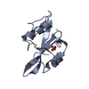
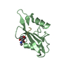
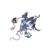
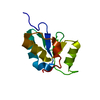
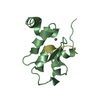
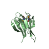
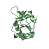
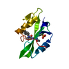
| 登録情報 | データベース: PDB / ID: 1nzl | ||||||
|---|---|---|---|---|---|---|---|
| タイトル | Crystal Structure of Src SH2 domain bound to doubly phosphorylated peptide PQpYEpYIPI | ||||||
 要素 要素 |
| ||||||
 キーワード キーワード | TRANSFERASE / SH2 domain / phosphotyrosine | ||||||
| 機能・相同性 |  機能・相同性情報 機能・相同性情報osteoclast development / progesterone receptor signaling pathway / negative regulation of intrinsic apoptotic signaling pathway / bone resorption / extrinsic component of cytoplasmic side of plasma membrane / negative regulation of extrinsic apoptotic signaling pathway / non-specific protein-tyrosine kinase / non-membrane spanning protein tyrosine kinase activity / epidermal growth factor receptor signaling pathway / cell adhesion ...osteoclast development / progesterone receptor signaling pathway / negative regulation of intrinsic apoptotic signaling pathway / bone resorption / extrinsic component of cytoplasmic side of plasma membrane / negative regulation of extrinsic apoptotic signaling pathway / non-specific protein-tyrosine kinase / non-membrane spanning protein tyrosine kinase activity / epidermal growth factor receptor signaling pathway / cell adhesion / phosphorylation / innate immune response / signaling receptor binding / ATP binding 類似検索 - 分子機能 | ||||||
| 生物種 |  Rous sarcoma virus (ラウス肉腫ウイルス) Rous sarcoma virus (ラウス肉腫ウイルス) | ||||||
| 手法 |  X線回折 / 分子置換 / 解像度: 1.9 Å X線回折 / 分子置換 / 解像度: 1.9 Å | ||||||
 データ登録者 データ登録者 | Lubman, O.Y. / Waksman, G. | ||||||
 引用 引用 | ジャーナル: J.Mol.Biol. / 年: 2003 タイトル: Structural and Thermodynamic Basis for the Interaction of the Src SH2 domain with the Activated form of the PDGF beta-receptor 著者: Lubman, O.Y. / Waksman, G. | ||||||
| 履歴 |
| ||||||
| Remark 600 | HETEROGEN The C8 and O5 atoms of the het hroup PG4 were not modelled and are not present in the coordinates. |
- 構造の表示
構造の表示
| 構造ビューア | 分子: MolmilJmol/JSmol |
|---|
- ダウンロードとリンク
-ダウンロード
| PDBx/mmCIF形式 | 1nzl.cif.gz | 59.9 KB | 表示 | PDBx/mmCIF形式 |
|---|---|---|---|---|
| PDB形式 | pdb1nzl.ent.gz | 42.2 KB | 表示 | PDB形式 |
| PDBx/mmJSON形式 | 1nzl.json.gz | ツリー表示 | PDBx/mmJSON形式 | |
| その他 |  その他のダウンロード その他のダウンロード |
-検証レポート
| 文書・要旨 | 1nzl_validation.pdf.gz | 455.8 KB | 表示 | wwPDB検証レポート |
|---|---|---|---|---|
| 文書・詳細版 | 1nzl_full_validation.pdf.gz | 458.7 KB | 表示 | |
| XML形式データ | 1nzl_validation.xml.gz | 12.7 KB | 表示 | |
| CIF形式データ | 1nzl_validation.cif.gz | 17 KB | 表示 | |
| アーカイブディレクトリ | https://data.pdbj.org/pub/pdb/validation_reports/nz/1nzlftp://data.pdbj.org/pub/pdb/validation_reports/nz/1nzl | HTTPS FTP |
-関連構造データ


-リンク
 PDBj
PDBj- 集合体
集合体
| 登録構造単位 | 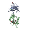
| ||||||||
|---|---|---|---|---|---|---|---|---|---|
| 1 | 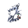
| ||||||||
| 2 | 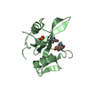
| ||||||||
| 単位格子 |
|
-要素
| #1: タンパク質 | 分子量: 11842.352 Da / 分子数: 2 / 断片: SH2 domain / 由来タイプ: 組換発現 由来: (組換発現) Rous sarcoma virus (strain Schmidt-Ruppin) (ラウス肉腫ウイルス)属: Alpharetrovirus / 生物種: Rous sarcoma virus / 株: Schmidt-Ruppin / 遺伝子: V-SRC / プラスミド: pET3 / 生物種 (発現宿主): Escherichia coli / 発現宿主:  #2: タンパク質・ペプチド | | 分子量: 1140.029 Da / 分子数: 1 / 由来タイプ: 合成 #3: 化合物 |   分子量: 35.453 Da / 分子数: 2 / 由来タイプ: 合成 / 式: Cl 分子量: 35.453 Da / 分子数: 2 / 由来タイプ: 合成 / 式: Cl#4: 化合物 | ChemComp-PG4 / |   分子量: 194.226 Da / 分子数: 1 / 由来タイプ: 合成 / 式: C8H18O5 / コメント: 沈殿剤*YM 分子量: 194.226 Da / 分子数: 1 / 由来タイプ: 合成 / 式: C8H18O5 / コメント: 沈殿剤*YM#5: 水 | ChemComp-HOH / |  分子量: 18.015 Da / 分子数: 163 / 由来タイプ: 天然 / 式: H2O 分子量: 18.015 Da / 分子数: 163 / 由来タイプ: 天然 / 式: H2O |
|---|
-実験情報
-実験
| 実験 | 手法: X線回折 / 使用した結晶の数: 1 |
|---|
- 試料調製
試料調製
| 結晶 | マシュー密度: 1.97 Å3/Da / 溶媒含有率: 37 % | |||||||||||||||||||||||||||||||||||
|---|---|---|---|---|---|---|---|---|---|---|---|---|---|---|---|---|---|---|---|---|---|---|---|---|---|---|---|---|---|---|---|---|---|---|---|---|
| 結晶化 | 温度: 296 K / 手法: 蒸気拡散法, ハンギングドロップ法 / pH: 8 詳細: PEG 4000,magnesium chloride, hepes, pH 8.0, VAPOR DIFFUSION, HANGING DROP, temperature 296K | |||||||||||||||||||||||||||||||||||
| 結晶化 | *PLUS | |||||||||||||||||||||||||||||||||||
| 溶液の組成 | *PLUS
|
-データ収集
| 回折 | 平均測定温度: 200 K |
|---|---|
| 放射光源 | 由来: 回転陽極 / タイプ: RIGAKU RU200 / 波長: 1.5418 Å |
| 放射 | モノクロメーター: graphite / プロトコル: SINGLE WAVELENGTH / 単色(M)・ラウエ(L): M / 散乱光タイプ: x-ray |
| 放射波長 | 波長: 1.5418 Å / 相対比: 1 |
| 反射 | 解像度: 1.9→30 Å / Num. obs: 17153 / % possible obs: 97.4 % / Observed criterion σ(F): 1 / Observed criterion σ(I): 2 / Biso Wilson estimate: 10.9 Å2 / Rsym value: 0.069 |
| 反射 | *PLUS 最低解像度: 30 Å / Num. measured all: 205689 / Rmerge(I) obs: 0.069 |
| 反射 シェル | *PLUS 最高解像度: 1.89 Å / 最低解像度: 1.96 Å / % possible obs: 93.2 % / Rmerge(I) obs: 0.163 |
- 解析
解析
| ソフトウェア |
| ||||||||||||||||||||||||||||||||||||||||||||||||||||||||||||||||||||||||||||||||
|---|---|---|---|---|---|---|---|---|---|---|---|---|---|---|---|---|---|---|---|---|---|---|---|---|---|---|---|---|---|---|---|---|---|---|---|---|---|---|---|---|---|---|---|---|---|---|---|---|---|---|---|---|---|---|---|---|---|---|---|---|---|---|---|---|---|---|---|---|---|---|---|---|---|---|---|---|---|---|---|---|---|
| 精密化 | 構造決定の手法: 分子置換 開始モデル: PDB ENTRY 1sps 解像度: 1.9→28.27 Å / Rfactor Rfree error: 0.007 / Isotropic thermal model: RESTRAINED / 交差検証法: THROUGHOUT / σ(F): 1 / 立体化学のターゲット値: Engh & Huber
| ||||||||||||||||||||||||||||||||||||||||||||||||||||||||||||||||||||||||||||||||
| 溶媒の処理 | 溶媒モデル: FLAT MODEL / Bsol: 47.7867 Å2 / ksol: 0.376681 e/Å3 | ||||||||||||||||||||||||||||||||||||||||||||||||||||||||||||||||||||||||||||||||
| 原子変位パラメータ | Biso mean: 20.1 Å2
| ||||||||||||||||||||||||||||||||||||||||||||||||||||||||||||||||||||||||||||||||
| Refine analyze |
| ||||||||||||||||||||||||||||||||||||||||||||||||||||||||||||||||||||||||||||||||
| 精密化ステップ | サイクル: LAST / 解像度: 1.9→28.27 Å
| ||||||||||||||||||||||||||||||||||||||||||||||||||||||||||||||||||||||||||||||||
| 拘束条件 |
| ||||||||||||||||||||||||||||||||||||||||||||||||||||||||||||||||||||||||||||||||
| LS精密化 シェル | 解像度: 1.9→2.02 Å / Rfactor Rfree error: 0.017 / Total num. of bins used: 6
| ||||||||||||||||||||||||||||||||||||||||||||||||||||||||||||||||||||||||||||||||
| Xplor file |
| ||||||||||||||||||||||||||||||||||||||||||||||||||||||||||||||||||||||||||||||||
| 精密化 | *PLUS 最高解像度: 1.9 Å / 最低解像度: 30 Å / Num. reflection obs: 16447 / % reflection Rfree: 10 % / Rfactor Rfree: 0.263 | ||||||||||||||||||||||||||||||||||||||||||||||||||||||||||||||||||||||||||||||||
| 溶媒の処理 | *PLUS | ||||||||||||||||||||||||||||||||||||||||||||||||||||||||||||||||||||||||||||||||
| 原子変位パラメータ | *PLUS | ||||||||||||||||||||||||||||||||||||||||||||||||||||||||||||||||||||||||||||||||
| 拘束条件 | *PLUS
|
