 Movie
Movie Controller

[English] 日本語
 Yorodumi
- PDB-1nxy: Crystal Structure of the complex between M182T mutant of TEM-1 an... -
+ Open data

- Basic information
| Entry | Database: PDB / ID: 1nxy | ||||||
|---|---|---|---|---|---|---|---|
| Title | Crystal Structure of the complex between M182T mutant of TEM-1 and a boronic acid inhibitor (SM2) | ||||||
 Components Components | Beta-lactamase TEM | ||||||
 Keywords Keywords | HYDROLASE / Antibiotic resistance / beta-lactamase / deacylation transition-state analog | ||||||
| Function / homology |  Function and homology information Function and homology informationAntimicrobial resistance / beta-lactam antibiotic catabolic process / beta-lactamase activity / beta-lactamase / response to antibiotic Similarity search - Function | ||||||
| Biological species |  | ||||||
| Method |  X-RAY DIFFRACTION / SYNCHROTRON / MOLECULAR REPLACEMENT / Resolution: 1.6 Å X-RAY DIFFRACTION / SYNCHROTRON / MOLECULAR REPLACEMENT / Resolution: 1.6 Å | ||||||
 Authors Authors | Wang, X. / Minasov, G. / Blazquez, J. / Caselli, E. / Prati, F. / Shoichet, B.K. | ||||||
 Citation Citation | Journal: Biochemistry / Year: 2003 Title: Recognition and Resistance in TEM beta-lactamase Authors: Wang, X. / Minasov, G. / Blazquez, J. / Caselli, E. / Prati, F. / Shoichet, B.K. | ||||||
| History |
|
- Structure visualization
| Structure viewer | Molecule: MolmilJmol/JSmol |
|---|
- Downloads & links
-Download
| PDBx/mmCIF format | 1nxy.cif.gz | 85 KB | Display | PDBx/mmCIF format |
|---|---|---|---|---|
| PDB format | pdb1nxy.ent.gz | 63.4 KB | Display | PDB format |
| PDBx/mmJSON format | 1nxy.json.gz | Tree view | PDBx/mmJSON format | |
| Others |  Other downloads Other downloads |
-Validation report
| Arichive directory | https://data.pdbj.org/pub/pdb/validation_reports/nx/1nxyftp://data.pdbj.org/pub/pdb/validation_reports/nx/1nxy | HTTPS FTP |
|---|
-Related structure data
| Related structure data |  1ny0C  1nymC  1nyyC  1jwpS S: Starting model for refinement C: citing same article ( |
|---|---|
| Similar structure data |


-Links
 PDBj
PDBj


- Assembly
| Deposited unit | 
| ||||||||
|---|---|---|---|---|---|---|---|---|---|
| 1 |
| ||||||||
| Unit cell |
|
-Components
| #1: Protein | Mass: 28911.904 Da / Num. of mol.: 1 / Mutation: M182T Source method: isolated from a genetically manipulated source Source: (gene. exp.) | ||||
|---|---|---|---|---|---|
| #2: Chemical | ChemComp-K /   Mass: 39.098 Da / Num. of mol.: 1 / Source method: obtained synthetically / Formula: K Mass: 39.098 Da / Num. of mol.: 1 / Source method: obtained synthetically / Formula: K | ||||
| #3: Chemical | 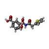  Mass: 319.141 Da / Num. of mol.: 2 / Source method: obtained synthetically / Formula: C14H14BNO5S Mass: 319.141 Da / Num. of mol.: 2 / Source method: obtained synthetically / Formula: C14H14BNO5S#4: Water | ChemComp-HOH / |  Mass: 18.015 Da / Num. of mol.: 447 / Source method: isolated from a natural source / Formula: H2O Mass: 18.015 Da / Num. of mol.: 447 / Source method: isolated from a natural source / Formula: H2OHas protein modification | Y | |
-Experimental details
-Experiment
| Experiment | Method: X-RAY DIFFRACTION / Number of used crystals: 1 |
|---|
- Sample preparation
| Crystal | Density Matthews: 2.32 Å3/Da / Density % sol: 46.94 % | ||||||||||||||||||||||||||||||
|---|---|---|---|---|---|---|---|---|---|---|---|---|---|---|---|---|---|---|---|---|---|---|---|---|---|---|---|---|---|---|---|
| Crystal grow | Temperature: 295 K / Method: vapor diffusion, hanging drop / pH: 8.3 Details: sodium-potassium buffer, pH 8.3, VAPOR DIFFUSION, HANGING DROP, temperature 295.0K | ||||||||||||||||||||||||||||||
| Crystal grow | *PLUS Method: vapor diffusion | ||||||||||||||||||||||||||||||
| Components of the solutions | *PLUS
|
-Data collection
| Diffraction | Mean temperature: 100 K |
|---|---|
| Diffraction source | Source: SYNCHROTRON / Site: APS  / Beamline: 5ID-B / Wavelength: 0.98012 Å / Beamline: 5ID-B / Wavelength: 0.98012 Å |
| Detector | Type: MARRESEARCH / Detector: CCD / Date: Nov 4, 2001 / Details: mirrors |
| Radiation | Monochromator: graphite / Protocol: SINGLE WAVELENGTH / Monochromatic (M) / Laue (L): M / Scattering type: x-ray |
| Radiation wavelength | Wavelength: 0.98012 Å / Relative weight: 1 |
| Reflection | Resolution: 1.6→15 Å / Num. all: 36330 / Num. obs: 36330 / % possible obs: 99.8 % / Observed criterion σ(F): 0 / Observed criterion σ(I): -3 / Redundancy: 6.1 % / Rmerge(I) obs: 0.053 / Net I/σ(I): 29.8 |
| Reflection shell | Resolution: 1.6→1.66 Å / Redundancy: 5.3 % / Rmerge(I) obs: 0.241 / Mean I/σ(I) obs: 7.2 / Num. unique all: 3568 / % possible all: 99.5 |
| Reflection | *PLUS Num. measured all: 220946 |
| Reflection shell | *PLUS % possible obs: 99.5 % / Num. unique obs: 3568 |
- Processing
| Software |
| ||||||||||||||||||||||||||||||||||||
|---|---|---|---|---|---|---|---|---|---|---|---|---|---|---|---|---|---|---|---|---|---|---|---|---|---|---|---|---|---|---|---|---|---|---|---|---|---|
| Refinement | Method to determine structure: MOLECULAR REPLACEMENT Starting model: PDB ENTRY 1JWP Resolution: 1.6→15 Å Isotropic thermal model: Isotropic, individual B values refined Cross valid method: THROUGHOUT / σ(F): 0 / σ(I): 0 / Stereochemistry target values: Engh & Huber Details: Maximum likelihood target for amplitudes was used in refinement
| ||||||||||||||||||||||||||||||||||||
| Displacement parameters | Biso mean: 15.8 Å2
| ||||||||||||||||||||||||||||||||||||
| Refine analyze |
| ||||||||||||||||||||||||||||||||||||
| Refinement step | Cycle: LAST / Resolution: 1.6→15 Å
| ||||||||||||||||||||||||||||||||||||
| Refine LS restraints |
| ||||||||||||||||||||||||||||||||||||
| LS refinement shell | Resolution: 1.6→1.66 Å
| ||||||||||||||||||||||||||||||||||||
| Xplor file |
| ||||||||||||||||||||||||||||||||||||
| Refinement | *PLUS % reflection Rfree: 10 % / Rfactor Rfree: 0.198 / Rfactor Rwork: 0.177 | ||||||||||||||||||||||||||||||||||||
| Solvent computation | *PLUS | ||||||||||||||||||||||||||||||||||||
| Displacement parameters | *PLUS | ||||||||||||||||||||||||||||||||||||
| Refine LS restraints | *PLUS
|
