 Movie
Movie Controller


+ Open data

- Basic information
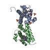
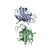
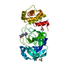
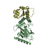
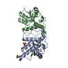
| Entry | Database: PDB / ID: 1nv9 | ||||||
|---|---|---|---|---|---|---|---|
| Title | HemK, apo structure | ||||||
 Components Components | hemK protein | ||||||
 Keywords Keywords | TRANSFERASE / Class I methyltransferase fold | ||||||
| Function / homology |  Function and homology information Function and homology informationpeptide chain release factor N5-glutamine methyltransferase / peptide chain release factor N(5)-glutamine methyltransferase activity / protein-glutamine N-methyltransferase activity / translational termination / methylation / nucleic acid binding Similarity search - Function | ||||||
| Biological species |   Thermotoga maritima (bacteria) Thermotoga maritima (bacteria) | ||||||
| Method |  X-RAY DIFFRACTION / SYNCHROTRON / MAD / Resolution: 2.356 Å X-RAY DIFFRACTION / SYNCHROTRON / MAD / Resolution: 2.356 Å | ||||||
 Authors Authors | Schubert, H.L. / Phillips, J.D. / Hill, C.P. | ||||||
 Citation Citation | Journal: Biochemistry / Year: 2003 Title: Structures along the Catalytic Pathway of PrmC/HemK, an N(5)-Glutamine AdoMet-Dependent Methyltransferase Authors: Schubert, H.L. / Phillips, J.D. / Hill, C.P. | ||||||
| History |
|
- Structure visualization
| Structure viewer | Molecule: MolmilJmol/JSmol |
|---|
- Downloads & links
-Download
| PDBx/mmCIF format | 1nv9.cif.gz | 66.6 KB | Display | PDBx/mmCIF format |
|---|---|---|---|---|
| PDB format | pdb1nv9.ent.gz | 48.4 KB | Display | PDB format |
| PDBx/mmJSON format | 1nv9.json.gz | Tree view | PDBx/mmJSON format | |
| Others |  Other downloads Other downloads |
-Validation report
| Arichive directory | https://data.pdbj.org/pub/pdb/validation_reports/nv/1nv9ftp://data.pdbj.org/pub/pdb/validation_reports/nv/1nv9 | HTTPS FTP |
|---|
-Related structure data
| Related structure data |  1nv8SC S: Starting model for refinement C: citing same article ( |
|---|---|
| Similar structure data |


-Links
 PDBj
PDBj- Assembly
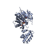
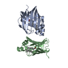
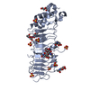
| Deposited unit | 
| ||||||||
|---|---|---|---|---|---|---|---|---|---|
| 1 |
| ||||||||
| Unit cell |
|
-Components
| #1: Protein | Mass: 31781.535 Da / Num. of mol.: 1 / Fragment: HemK Source method: isolated from a genetically manipulated source Source: (gene. exp.) Thermotoga maritima (bacteria) / Gene: HemK / Plasmid: pET14b / Production host: References: UniProt: Q9WYV8, Transferases; Transferring one-carbon groups; Methyltransferases |
|---|---|
| #2: Chemical | ChemComp-SAH /   Type: L-peptide linking / Mass: 384.411 Da / Num. of mol.: 1 / Source method: obtained synthetically / Formula: C14H20N6O5S Type: L-peptide linking / Mass: 384.411 Da / Num. of mol.: 1 / Source method: obtained synthetically / Formula: C14H20N6O5S |
| #3: Water | ChemComp-HOH /  Mass: 18.015 Da / Num. of mol.: 20 / Source method: isolated from a natural source / Formula: H2O Mass: 18.015 Da / Num. of mol.: 20 / Source method: isolated from a natural source / Formula: H2O |
-Experimental details
-Experiment
| Experiment | Method: X-RAY DIFFRACTION / Number of used crystals: 1 |
|---|
- Sample preparation
| Crystal | Density Matthews: 2.46 Å3/Da / Density % sol: 49.91 % | ||||||||||||||||||||||||
|---|---|---|---|---|---|---|---|---|---|---|---|---|---|---|---|---|---|---|---|---|---|---|---|---|---|
| Crystal grow | Temperature: 292 K / Method: vapor diffusion, hanging drop / pH: 5.5 Details: PEG3000, Citrate, pH 5.5, VAPOR DIFFUSION, HANGING DROP, temperature 19K | ||||||||||||||||||||||||
| Crystal grow | *PLUS Method: vapor diffusion, hanging drop | ||||||||||||||||||||||||
| Components of the solutions | *PLUS
|
-Data collection
| Diffraction | Mean temperature: 100 K |
|---|---|
| Diffraction source | Source: SYNCHROTRON / Site: SSRL  / Beamline: BL9-2 / Wavelength: 0.97 Å / Beamline: BL9-2 / Wavelength: 0.97 Å |
| Detector | Type: ADSC QUANTUM 4 / Detector: CCD / Date: Mar 30, 2002 |
| Radiation | Protocol: SINGLE WAVELENGTH / Monochromatic (M) / Laue (L): M / Scattering type: x-ray |
| Radiation wavelength | Wavelength: 0.97 Å / Relative weight: 1 |
| Reflection | Resolution: 2.35→50 Å / Num. all: 13364 / Num. obs: 13340 / % possible obs: 97.7 % / Observed criterion σ(F): 0 / Observed criterion σ(I): 0 / Rmerge(I) obs: 0.039 / Net I/σ(I): 20 |
| Reflection shell | Resolution: 2.35→2.43 Å / Rmerge(I) obs: 0.404 / Mean I/σ(I) obs: 2.3 / % possible all: 79.4 |
| Reflection | *PLUS Lowest resolution: 20 Å / Num. obs: 24661 / Num. measured all: 100547 |
| Reflection shell | *PLUS % possible obs: 79.4 % |
- Processing
| Software |
| ||||||||||||||||||||||||||||||||||||||||||||||||||||||||||||||||||||||||||||||||||||||||||||||||||||||||||||||
|---|---|---|---|---|---|---|---|---|---|---|---|---|---|---|---|---|---|---|---|---|---|---|---|---|---|---|---|---|---|---|---|---|---|---|---|---|---|---|---|---|---|---|---|---|---|---|---|---|---|---|---|---|---|---|---|---|---|---|---|---|---|---|---|---|---|---|---|---|---|---|---|---|---|---|---|---|---|---|---|---|---|---|---|---|---|---|---|---|---|---|---|---|---|---|---|---|---|---|---|---|---|---|---|---|---|---|---|---|---|---|---|
| Refinement | Method to determine structure: MAD Starting model: PDB ENTRY 1NV8 Resolution: 2.356→50 Å / Cor.coef. Fo:Fc: 0.922 / Cor.coef. Fo:Fc free: 0.892 / SU B: 13.479 / SU ML: 0.326 / Cross valid method: THROUGHOUT / σ(F): 0 / ESU R: 0.41 / ESU R Free: 0.296 / Stereochemistry target values: MAXIMUM LIKELIHOOD / Details: HYDROGENS HAVE BEEN ADDED IN THE RIDING POSITIONS
| ||||||||||||||||||||||||||||||||||||||||||||||||||||||||||||||||||||||||||||||||||||||||||||||||||||||||||||||
| Solvent computation | Ion probe radii: 0.8 Å / Shrinkage radii: 0.8 Å / VDW probe radii: 1.4 Å / Solvent model: BABINET MODEL WITH MASK | ||||||||||||||||||||||||||||||||||||||||||||||||||||||||||||||||||||||||||||||||||||||||||||||||||||||||||||||
| Displacement parameters | Biso mean: 41.043 Å2
| ||||||||||||||||||||||||||||||||||||||||||||||||||||||||||||||||||||||||||||||||||||||||||||||||||||||||||||||
| Refinement step | Cycle: LAST / Resolution: 2.356→50 Å
| ||||||||||||||||||||||||||||||||||||||||||||||||||||||||||||||||||||||||||||||||||||||||||||||||||||||||||||||
| Refine LS restraints |
| ||||||||||||||||||||||||||||||||||||||||||||||||||||||||||||||||||||||||||||||||||||||||||||||||||||||||||||||
| LS refinement shell | Resolution: 2.356→2.417 Å / Total num. of bins used: 20 /
| ||||||||||||||||||||||||||||||||||||||||||||||||||||||||||||||||||||||||||||||||||||||||||||||||||||||||||||||
| Software | *PLUS Version: 5 / Classification: refinement | ||||||||||||||||||||||||||||||||||||||||||||||||||||||||||||||||||||||||||||||||||||||||||||||||||||||||||||||
| Refinement | *PLUS Highest resolution: 2.35 Å / Lowest resolution: 20 Å / % reflection Rfree: 5 % / Rfactor Rfree: 0.3 / Rfactor Rwork: 0.238 | ||||||||||||||||||||||||||||||||||||||||||||||||||||||||||||||||||||||||||||||||||||||||||||||||||||||||||||||
| Solvent computation | *PLUS | ||||||||||||||||||||||||||||||||||||||||||||||||||||||||||||||||||||||||||||||||||||||||||||||||||||||||||||||
| Displacement parameters | *PLUS | ||||||||||||||||||||||||||||||||||||||||||||||||||||||||||||||||||||||||||||||||||||||||||||||||||||||||||||||
| Refine LS restraints | *PLUS
|
