Resolution: 2.8→36.09 Å / Cor.coef. Fo:Fc: 0.954 / Cor.coef. Fo:Fc free: 0.901 / SU B: 42.973 / SU ML: 0.375 / TLS residual ADP flag: LIKELY RESIDUAL / Cross valid method: THROUGHOUT / ESU R Free: 0.416 / Stereochemistry target values: MAXIMUM LIKELIHOOD Details: HYDROGENS HAVE BEEN ADDED IN THE RIDING POSITIONS. DUE TO LACK OF DENSITY, THE FOLLOWING REGIONS WERE NOT MODELED: A/B1-11, A207-211, B205-211. SAM MODELED BASED ON RELATED STRUCTURE (1NV8)
Rfactor
Num. reflection
% reflection
Selection details
Rfree
0.27281
801
5.1 %
RANDOM
Rwork
0.20824
-
-
-
obs
0.21156
15024
95.94 %
-
Solvent computation
Ion probe radii: 0.8 Å / Shrinkage radii: 0.8 Å / VDW probe radii: 1.2 Å / Solvent model: MASK
Displacement parameters
Biso mean: 85.35 Å2
Baniso -1
Baniso -2
Baniso -3
1-
5.5 Å2
0 Å2
-1.54 Å2
2-
-
-7.5 Å2
0 Å2
3-
-
-
0.98 Å2
Refinement step
Cycle: LAST / Resolution: 2.8→36.09 Å
Protein
Nucleic acid
Ligand
Solvent
Total
Num. atoms
4116
0
54
7
4177
Refine LS restraints
Refine-ID
Type
Dev ideal
Dev ideal target
Number
X-RAY DIFFRACTION
r_bond_refined_d
0.014
0.022
4246
X-RAY DIFFRACTION
r_bond_other_d
0.002
0.02
4005
X-RAY DIFFRACTION
r_angle_refined_deg
1.507
2.002
5728
X-RAY DIFFRACTION
r_angle_other_deg
0.78
3
9256
X-RAY DIFFRACTION
r_dihedral_angle_1_deg
7.197
5
526
X-RAY DIFFRACTION
r_dihedral_angle_2_deg
30.817
23.146
178
X-RAY DIFFRACTION
r_dihedral_angle_3_deg
17.375
15
744
X-RAY DIFFRACTION
r_dihedral_angle_4_deg
16.09
15
33
X-RAY DIFFRACTION
r_chiral_restr
0.071
0.2
654
X-RAY DIFFRACTION
r_gen_planes_refined
0.005
0.02
4658
X-RAY DIFFRACTION
r_gen_planes_other
0.002
0.02
891
X-RAY DIFFRACTION
r_nbd_refined
0.23
0.2
988
X-RAY DIFFRACTION
r_nbd_other
0.194
0.2
4203
X-RAY DIFFRACTION
r_nbtor_other
0.089
0.2
2605
X-RAY DIFFRACTION
r_xyhbond_nbd_refined
0.183
0.2
105
X-RAY DIFFRACTION
r_symmetry_vdw_refined
0.232
0.2
20
X-RAY DIFFRACTION
r_symmetry_vdw_other
0.22
0.2
61
X-RAY DIFFRACTION
r_symmetry_hbond_refined
0.352
0.2
1
X-RAY DIFFRACTION
r_mcbond_it
0.995
3
2689
X-RAY DIFFRACTION
r_mcbond_other
0.383
3
1094
X-RAY DIFFRACTION
r_mcangle_it
1.394
5
4219
X-RAY DIFFRACTION
r_scbond_it
3.285
8
1736
X-RAY DIFFRACTION
r_scangle_it
4.495
11
1509
X-RAY DIFFRACTION
r_nbtor_refined
0.193
0.2
2117
Refine LS restraints NCS
Dom-ID: 1 / Auth asym-ID: A / Refine-ID: X-RAY DIFFRACTION
Ens-ID
Number
Type
Rms dev position (Å)
Weight position
1
435
tightpositional
0.03
0.05
1
688
mediumpositional
0.31
0.5
1
435
tightthermal
0.11
0.5
1
688
mediumthermal
0.58
2
2
1117
tightpositional
0.04
0.05
2
1749
mediumpositional
0.36
0.5
2
1117
tightthermal
0.14
0.5
2
1749
mediumthermal
0.76
2
LS refinement shell
Resolution: 2.8→2.873 Å / Total num. of bins used: 20
Rfactor
Num. reflection
% reflection
Rfree
0.339
53
6.02 %
Rwork
0.346
827
-
obs
-
-
72.79 %
Refinement TLS params.
Method: refined / Refine-ID: X-RAY DIFFRACTION
ID
L11 (°2)
L12 (°2)
L13 (°2)
L22 (°2)
L23 (°2)
L33 (°2)
S11 (Å °)
S12 (Å °)
S13 (Å °)
S21 (Å °)
S22 (Å °)
S23 (Å °)
S31 (Å °)
S32 (Å °)
S33 (Å °)
T11 (Å2)
T12 (Å2)
T13 (Å2)
T22 (Å2)
T23 (Å2)
T33 (Å2)
Origin x (Å)
Origin y (Å)
Origin z (Å)
1
6.0832
0.1555
-2.3971
11.4257
1.8002
9.7491
0.5264
0.45
0.3394
-0.6257
-0.1156
-0.2067
0.0728
-0.1706
-0.4109
-0.0491
0.0373
-0.2921
-0.0124
0.1524
-0.2841
6.9881
11.9553
9.8928
2
3.9033
0.1722
1.4396
5.8365
-1.3302
8.0099
0.1494
-0.2047
-0.0598
0.1087
0.2699
0.7238
0.1546
-1.1116
-0.4193
-0.4297
-0.0136
-0.0507
-0.3434
0.0767
-0.3085
10.1764
-3.239
38.6992
3
12.3897
-4.567
2.9977
28.3096
2.6181
29.8229
0.805
-0.5403
1.2476
1.317
-1.2841
0.1522
-2.4636
-1.783
0.4791
0.1358
-0.0728
0.2074
-0.0752
0.1388
0.2046
5.4521
-28.0856
15.6415
4
5.7876
0.3821
-0.8862
8.9155
-1.3277
5.2313
-0.1632
0.7347
0.2623
-0.0645
0.5323
1.2155
0.0553
-0.5838
-0.369
-0.3091
-0.0006
0.0578
-0.3599
0.0997
-0.0858
26.104
-13.4976
-5.3668
Refinement TLS group
Refine-ID: X-RAY DIFFRACTION / Selection: ALL
ID
Refine TLS-ID
Auth asym-ID
Label asym-ID
Auth seq-ID
Label seq-ID
1
1
A
A
12 - 85
24 - 97
2
2
A
A
86 - 282
98 - 294
3
3
B
B
12 - 85
24 - 97
4
4
B
B
86 - 282
98 - 294
+
About Yorodumi
-
News
-
Feb 9, 2022. New format data for meta-information of EMDB entries
New format data for meta-information of EMDB entries
Version 3 of the EMDB header file is now the official format.
The previous official version 1.9 will be removed from the archive.
In the structure databanks used in Yorodumi, some data are registered as the other names, "COVID-19 virus" and "2019-nCoV". Here are the details of the virus and the list of structure data.
Jan 31, 2019. EMDB accession codes are about to change! (news from PDBe EMDB page)
EMDB accession codes are about to change! (news from PDBe EMDB page)
The allocation of 4 digits for EMDB accession codes will soon come to an end. Whilst these codes will remain in use, new EMDB accession codes will include an additional digit and will expand incrementally as the available range of codes is exhausted. The current 4-digit format prefixed with “EMD-” (i.e. EMD-XXXX) will advance to a 5-digit format (i.e. EMD-XXXXX), and so on. It is currently estimated that the 4-digit codes will be depleted around Spring 2019, at which point the 5-digit format will come into force.
The EM Navigator/Yorodumi systems omit the EMD- prefix.
Related info.:Q: What is EMD? / ID/Accession-code notation in Yorodumi/EM Navigator
Yorodumi is a browser for structure data from EMDB, PDB, SASBDB, etc.
This page is also the successor to EM Navigator detail page, and also detail information page/front-end page for Omokage search.
The word "yorodu" (or yorozu) is an old Japanese word meaning "ten thousand". "mi" (miru) is to see.
Related info.:EMDB / PDB / SASBDB / Comparison of 3 databanks / Yorodumi Search / Aug 31, 2016. New EM Navigator & Yorodumi / Yorodumi Papers / Jmol/JSmol / Function and homology information / Changes in new EM Navigator and Yorodumi
 Movie
Movie Controller

 Yorodumi
 Open data
 Basic information
 Components
 Keywords
 Function and homology information
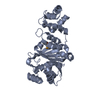
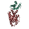
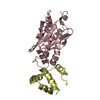
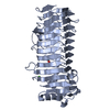
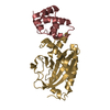
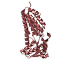
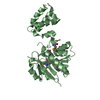
 Thermotoga maritima (bacteria)
 X-RAY DIFFRACTION /
  Authors
 Citation
 Structure visualization
 Downloads & links
 Other downloads


 PDBj
PDBj
 Assembly

 Mass: 398.437 Da / Num. of mol.: 2 / Source method: obtained synthetically / Formula: C15H22N6O5S
 Mass: 18.015 Da / Num. of mol.: 7 / Source method: isolated from a natural source / Formula: H2O
 Sample preparation
 / Beamline: 8.2.1 / Wavelength: 0.9796
  Processing