 Movie
Movie Controller

[English] 日本語
 Yorodumi
- PDB-1ns6: The 2.1A Structure of Horse (alpha hemichrome/beta met) Hemoglobi... -
+ Open data

- Basic information
| Entry | Database: PDB / ID: 1ns6 | ||||||
|---|---|---|---|---|---|---|---|
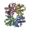
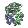
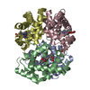
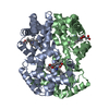
| Title | The 2.1A Structure of Horse (alpha hemichrome/beta met) Hemoglobin at pH 5.4 | ||||||
 Components Components |
| ||||||
 Keywords Keywords | OXYGEN STORAGE/TRANSPORT / hemichrome / bisimidazole alpha heme / aquomet beta heme / globin fold / OXYGEN STORAGE-TRANSPORT COMPLEX | ||||||
| Function / homology |  Function and homology information Function and homology informationhemoglobin alpha binding / haptoglobin-hemoglobin complex / hemoglobin complex / oxygen transport / erythrocyte development / oxygen carrier activity / oxygen binding / iron ion binding / heme binding / metal ion binding Similarity search - Function | ||||||
| Biological species |  | ||||||
| Method |  X-RAY DIFFRACTION / MOLECULAR REPLACEMENT / Resolution: 2.05 Å X-RAY DIFFRACTION / MOLECULAR REPLACEMENT / Resolution: 2.05 Å | ||||||
 Authors Authors | Robinson, V.L. / Smith, B.B. / Arnone, A. | ||||||
 Citation Citation | Journal: Biochemistry / Year: 2003 Title: A pH-Dependent Aquomet-to-Hemichrome Transition in Crystalline Horse Methemoglobin Authors: Robinson, V.L. / Smith, B.B. / Arnone, A. #1: Journal: Proc.R.Soc.London,Ser.B / Year: 1947Title: An X-Ray Study of Horse Methaemoglobin. I Authors: Boyes-Watson, J. / Davidson, E. / Perutz, M.F. #2: Journal: Proc.R.Soc.London,Ser.B / Year: 1954Title: The Structure of Haemoglobin. III. Direct Determination of the Molecular Transform Authors: Perutz, M.F. | ||||||
| History |
|
- Structure visualization
| Structure viewer | Molecule: MolmilJmol/JSmol |
|---|
- Downloads & links
-Download
| PDBx/mmCIF format | 1ns6.cif.gz | 71.5 KB | Display | PDBx/mmCIF format |
|---|---|---|---|---|
| PDB format | pdb1ns6.ent.gz | 52.6 KB | Display | PDB format |
| PDBx/mmJSON format | 1ns6.json.gz | Tree view | PDBx/mmJSON format | |
| Others |  Other downloads Other downloads |
-Validation report
| Arichive directory | https://data.pdbj.org/pub/pdb/validation_reports/ns/1ns6ftp://data.pdbj.org/pub/pdb/validation_reports/ns/1ns6 | HTTPS FTP |
|---|
-Related structure data
| Related structure data | 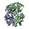 1ns9C 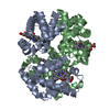 1g0bS C: citing same article ( S: Starting model for refinement |
|---|---|
| Similar structure data |

-Links
 PDBj
PDBj


- Assembly
| Deposited unit | 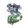
| ||||||||||
|---|---|---|---|---|---|---|---|---|---|---|---|
| 1 | 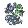
| ||||||||||
| Unit cell |
| ||||||||||
| Details | The biological assembly (tetramer) is generated from the dimer in the asymetric unit by the operation: -x, y, -z. |
-Components
| #1: Protein | Mass: 15138.280 Da / Num. of mol.: 1 / Fragment: alpha subunit / Source method: isolated from a natural source / Details: pH 5.4 Hemichrome Structure / Source: (natural) | ||
|---|---|---|---|
| #2: Protein | Mass: 16032.274 Da / Num. of mol.: 1 / Fragment: beta subunit / Source method: isolated from a natural source / Details: pH 5.4 Hemichrome Structure / Source: (natural) | ||
| #3: Chemical |   Mass: 616.487 Da / Num. of mol.: 2 / Source method: obtained synthetically / Formula: C34H32FeN4O4 Mass: 616.487 Da / Num. of mol.: 2 / Source method: obtained synthetically / Formula: C34H32FeN4O4#4: Water | ChemComp-HOH / |  Mass: 18.015 Da / Num. of mol.: 84 / Source method: isolated from a natural source / Formula: H2O Mass: 18.015 Da / Num. of mol.: 84 / Source method: isolated from a natural source / Formula: H2O |
-Experimental details
-Experiment
| Experiment | Method: X-RAY DIFFRACTION / Number of used crystals: 1 |
|---|
- Sample preparation
| Crystal | Density Matthews: 2.76 Å3/Da / Density % sol: 55.1 % | ||||||||||||||||||||||||
|---|---|---|---|---|---|---|---|---|---|---|---|---|---|---|---|---|---|---|---|---|---|---|---|---|---|
| Crystal grow | Temperature: 298 K / Method: vapor diffusion / pH: 5.4 Details: 1.5M ammonium sulfate, 0.35M ammonium phosphate then soaked to pH 5.4 in solutions of 3.35 M ammonium sulfate buffered with 0.2M ammonium phosphate, VAPOR DIFFUSION, temperature 298K | ||||||||||||||||||||||||
| Crystal grow | *PLUS pH: 7.1 / Method: batch method | ||||||||||||||||||||||||
| Components of the solutions | *PLUS
|
-Data collection
| Diffraction | Mean temperature: 298 K |
|---|---|
| Diffraction source | Source: ROTATING ANODE / Type: RIGAKU RU200 / Wavelength: 1.5418 Å |
| Detector | Type: UCSD MARK II / Detector: AREA DETECTOR / Date: Sep 15, 1997 |
| Radiation | Monochromator: graphite / Protocol: SINGLE WAVELENGTH / Monochromatic (M) / Laue (L): M / Scattering type: x-ray |
| Radiation wavelength | Wavelength: 1.5418 Å / Relative weight: 1 |
| Reflection | Resolution: 2.04→30 Å / Num. obs: 21884 / % possible obs: 97.2 % / Observed criterion σ(I): 2 / Rmerge(I) obs: 0.053 |
| Reflection | *PLUS Highest resolution: 2.07 Å / Num. obs: 21680 / Num. measured all: 125115 |
| Reflection shell | *PLUS Highest resolution: 2.07 Å / Lowest resolution: 2.23 Å / % possible obs: 87.7 % / Rmerge(I) obs: 0.123 / Mean I/σ(I) obs: 4.5 |
- Processing
| Software |
| ||||||||||||||||||||||||||||||||||||||||||||||||||||||||||||||||||||||||||||||||||||||||||||||||||||
|---|---|---|---|---|---|---|---|---|---|---|---|---|---|---|---|---|---|---|---|---|---|---|---|---|---|---|---|---|---|---|---|---|---|---|---|---|---|---|---|---|---|---|---|---|---|---|---|---|---|---|---|---|---|---|---|---|---|---|---|---|---|---|---|---|---|---|---|---|---|---|---|---|---|---|---|---|---|---|---|---|---|---|---|---|---|---|---|---|---|---|---|---|---|---|---|---|---|---|---|---|---|
| Refinement | Method to determine structure: MOLECULAR REPLACEMENT Starting model: PDB entry 1G0B Resolution: 2.05→54.23 Å / Cor.coef. Fo:Fc: 0.953 / SU B: 4.946 / SU ML: 0.136 / σ(F): 2 / ESU R: 0.149 / Stereochemistry target values: MAXIMUM LIKELIHOOD / Details: HYDROGENS HAVE BEEN ADDED IN THE RIDING POSITIONS
| ||||||||||||||||||||||||||||||||||||||||||||||||||||||||||||||||||||||||||||||||||||||||||||||||||||
| Solvent computation | Ion probe radii: 0.8 Å / Shrinkage radii: 0.8 Å / VDW probe radii: 1.4 Å / Solvent model: BABINET MODEL WITH MASK | ||||||||||||||||||||||||||||||||||||||||||||||||||||||||||||||||||||||||||||||||||||||||||||||||||||
| Displacement parameters | Biso mean: 18.383 Å2
| ||||||||||||||||||||||||||||||||||||||||||||||||||||||||||||||||||||||||||||||||||||||||||||||||||||
| Refinement step | Cycle: LAST / Resolution: 2.05→54.23 Å
| ||||||||||||||||||||||||||||||||||||||||||||||||||||||||||||||||||||||||||||||||||||||||||||||||||||
| Refine LS restraints |
| ||||||||||||||||||||||||||||||||||||||||||||||||||||||||||||||||||||||||||||||||||||||||||||||||||||
| LS refinement shell | Resolution: 2.05→2.103 Å / Num. reflection Rfree: 0 / Num. reflection Rwork: 838 / Total num. of bins used: 20 | ||||||||||||||||||||||||||||||||||||||||||||||||||||||||||||||||||||||||||||||||||||||||||||||||||||
| Software | *PLUS Version: 5 / Classification: refinement | ||||||||||||||||||||||||||||||||||||||||||||||||||||||||||||||||||||||||||||||||||||||||||||||||||||
| Refinement | *PLUS Highest resolution: 2.07 Å / Rfactor Rfree: 0.194 / Rfactor Rwork: 0.162 | ||||||||||||||||||||||||||||||||||||||||||||||||||||||||||||||||||||||||||||||||||||||||||||||||||||
| Solvent computation | *PLUS | ||||||||||||||||||||||||||||||||||||||||||||||||||||||||||||||||||||||||||||||||||||||||||||||||||||
| Displacement parameters | *PLUS | ||||||||||||||||||||||||||||||||||||||||||||||||||||||||||||||||||||||||||||||||||||||||||||||||||||
| Refine LS restraints | *PLUS
| ||||||||||||||||||||||||||||||||||||||||||||||||||||||||||||||||||||||||||||||||||||||||||||||||||||
| LS refinement shell | *PLUS Highest resolution: 2.07 Å / Lowest resolution: 2.23 Å |
