 Movie
Movie Controller

[English] 日本語
 Yorodumi
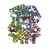
- PDB-1fhj: CRYSTAL STRUCTURE OF AQUOMET HEMOGLOBIN-I OF THE MANED WOLF (CHRY... -
+ Open data

- Basic information
| Entry | Database: PDB / ID: 1fhj | ||||||
|---|---|---|---|---|---|---|---|
| Title | CRYSTAL STRUCTURE OF AQUOMET HEMOGLOBIN-I OF THE MANED WOLF (CHRYSOCYON BRACHYURUS) AT 2.0 RESOLUTION. | ||||||
 Components Components |
| ||||||
 Keywords Keywords | OXYGEN STORAGE/TRANSPORT / maned wolf / hemoglobin / metahemoglobin / OXYGEN STORAGE-TRANSPORT COMPLEX | ||||||
| Function / homology |  Function and homology information Function and homology informationhaptoglobin binding / organic acid binding / hemoglobin alpha binding / haptoglobin-hemoglobin complex / hemoglobin complex / hydrogen peroxide catabolic process / oxygen carrier activity / peroxidase activity / oxygen binding / blood microparticle ...haptoglobin binding / organic acid binding / hemoglobin alpha binding / haptoglobin-hemoglobin complex / hemoglobin complex / hydrogen peroxide catabolic process / oxygen carrier activity / peroxidase activity / oxygen binding / blood microparticle / iron ion binding / heme binding / metal ion binding Similarity search - Function | ||||||
| Biological species |  Chrysocyon brachyurus (maned wolf) Chrysocyon brachyurus (maned wolf) | ||||||
| Method |  X-RAY DIFFRACTION / SYNCHROTRON / MOLECULAR REPLACEMENT / Resolution: 1.8 Å X-RAY DIFFRACTION / SYNCHROTRON / MOLECULAR REPLACEMENT / Resolution: 1.8 Å | ||||||
 Authors Authors | Fadel, V. / de Azevedo, W.F. | ||||||
 Citation Citation | Journal: Protein Pept.Lett. / Year: 2003 Title: Crystal structure of hemoglobin from the maned wolf (Chrysocyon brachyurus) using synchrotron radiation. Authors: Fadel, V. / Canduri, F. / Olivieri, J.R. / Smarra, A.L. / Colombo, M.F. / Bonilla-Rodriguez, G.O. / de Azevedo, W.F. #1: Journal: Acta Crystallogr.,Sect.D / Year: 1999Title: Crystallization, Preliminary X-ray Diffraction Analysis and Patterson Search of Oxyhemoglobin I from the Wolf (Chrysocyon brachiurus) Authors: Smarra, A.L. / Fadel, V. / Dellamano, M. / Olivieri, J.R. / de Azevedo Jr., W.F. / Bonilla-Rodriguez, G.O. | ||||||
| History |
|
- Structure visualization
| Structure viewer | Molecule: MolmilJmol/JSmol |
|---|
- Downloads & links
-Download
| PDBx/mmCIF format | 1fhj.cif.gz | 132.8 KB | Display | PDBx/mmCIF format |
|---|---|---|---|---|
| PDB format | pdb1fhj.ent.gz | 104.2 KB | Display | PDB format |
| PDBx/mmJSON format | 1fhj.json.gz | Tree view | PDBx/mmJSON format | |
| Others |  Other downloads Other downloads |
-Validation report
| Arichive directory | https://data.pdbj.org/pub/pdb/validation_reports/fh/1fhjftp://data.pdbj.org/pub/pdb/validation_reports/fh/1fhj | HTTPS FTP |
|---|
-Related structure data
| Related structure data | 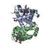 1hhoS S: Starting model for refinement |
|---|---|
| Similar structure data |
-Links
 PDBj
PDBj


- Assembly
| Deposited unit | 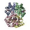
| ||||||||
|---|---|---|---|---|---|---|---|---|---|
| 1 |
| ||||||||
| Unit cell |
| ||||||||
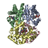
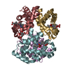
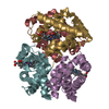
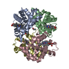
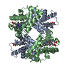
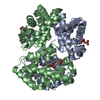
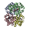
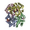
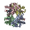
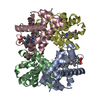
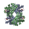
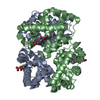
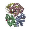
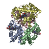
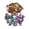
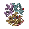
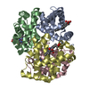
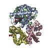
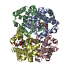
| Details | The asymmetric unit has a complete tetramer formed by two alpha chains and two beta chains. |
-Components
| #1: Protein | Mass: 15270.329 Da / Num. of mol.: 2 / Source method: isolated from a natural source / Source: (natural) Chrysocyon brachyurus (maned wolf) / References: UniProt: P01952, UniProt: P60523*PLUS#2: Protein | Mass: 16020.366 Da / Num. of mol.: 2 / Source method: isolated from a natural source / Source: (natural) Chrysocyon brachyurus (maned wolf) / References: UniProt: P02056, UniProt: P60526*PLUS#3: Chemical | ChemComp-HEM /   Mass: 616.487 Da / Num. of mol.: 4 / Source method: obtained synthetically / Formula: C34H32FeN4O4 Mass: 616.487 Da / Num. of mol.: 4 / Source method: obtained synthetically / Formula: C34H32FeN4O4#4: Water | ChemComp-HOH / |  Mass: 18.015 Da / Num. of mol.: 308 / Source method: isolated from a natural source / Formula: H2O Mass: 18.015 Da / Num. of mol.: 308 / Source method: isolated from a natural source / Formula: H2O |
|---|
-Experimental details
-Experiment
| Experiment | Method: X-RAY DIFFRACTION / Number of used crystals: 1 |
|---|
- Sample preparation
| Crystal | Density Matthews: 2.53 Å3/Da / Density % sol: 51.44 % |
|---|---|
| Crystal grow | Temperature: 295 K / Method: vapor diffusion / Details: PEG 1500 30%, VAPOR DIFFUSION, temperature 295K |
-Data collection
| Diffraction | Mean temperature: 300 K |
|---|---|
| Diffraction source | Source: SYNCHROTRON / Site: LNLS  / Beamline: D03B-MX1 / Wavelength: 1.37 / Beamline: D03B-MX1 / Wavelength: 1.37 |
| Detector | Type: MARRESEARCH / Detector: IMAGE PLATE / Date: Aug 27, 1998 |
| Radiation | Protocol: SINGLE WAVELENGTH / Monochromatic (M) / Laue (L): M / Scattering type: x-ray |
| Radiation wavelength | Wavelength: 1.37 Å / Relative weight: 1 |
| Reflection | Resolution: 1.864→72.548 Å / Num. all: 53941 / Num. obs: 50240 / % possible obs: 93.1 % / Observed criterion σ(I): 1 / Redundancy: 2.62 % / Rmerge(I) obs: 0.05 / Net I/σ(I): 9.4 |
| Reflection shell | Resolution: 1.86→1.9 Å / Redundancy: 2.38 % / Rmerge(I) obs: 0.583 / Num. unique all: 2799 / % possible all: 78.7 |
- Processing
| Software |
| |||||||||||||||||||||||||
|---|---|---|---|---|---|---|---|---|---|---|---|---|---|---|---|---|---|---|---|---|---|---|---|---|---|---|
| Refinement | Method to determine structure: MOLECULAR REPLACEMENT Starting model: HUMAN OXYHEMOGLOBIN pdb id 1HHO Resolution: 1.8→10 Å / σ(F): 2 / σ(I): 0 Details: Refmac was also used for refinement. THE SIMULLATED ANNEALING METHOD (X-PLOR) AND MAXIMUM LIKELIHOOD METHOD (REFMAC) WAS EMPLOYED. Data was used to 1.8 angstroms, but with low completeness.
| |||||||||||||||||||||||||
| Refinement step | Cycle: LAST / Resolution: 1.8→10 Å
| |||||||||||||||||||||||||
| Refine LS restraints |
|
