 Movie
Movie Controller

[English] 日本語
 Yorodumi
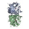
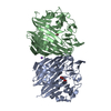
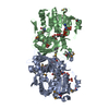
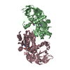
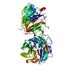
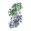
- PDB-1ns2: Crystal structure of galactose mutarotase from Lactococcus lactis... -
+ Open data

- Basic information
| Entry | Database: PDB / ID: 1ns2 | ||||||
|---|---|---|---|---|---|---|---|
| Title | Crystal structure of galactose mutarotase from Lactococcus lactis mutant E304A complexed with galactose | ||||||
 Components Components | GALACTOSE MUTAROTASE | ||||||
 Keywords Keywords | ISOMERASE / MUTAROTASE / EPIMERASE / GALACTOSE METABOLISM | ||||||
| Function / homology |  Function and homology information Function and homology informationaldose 1-epimerase / aldose 1-epimerase activity / beta-D-galactose catabolic process via UDP-galactose, Leloir pathway / glucose metabolic process / carbohydrate binding / cytoplasm Similarity search - Function | ||||||
| Biological species |  Lactococcus lactis (lactic acid bacteria) Lactococcus lactis (lactic acid bacteria) | ||||||
| Method |  X-RAY DIFFRACTION / FOURIER SYNTHESIS / Resolution: 1.95 Å X-RAY DIFFRACTION / FOURIER SYNTHESIS / Resolution: 1.95 Å | ||||||
 Authors Authors | Holden, H.M. / Thoden, J.B. | ||||||
 Citation Citation | Journal: Protein Sci. / Year: 2003 Title: The Catalytic Mechanism of Galactose Mutarotase Authors: Thoden, J.B. / Kim, J. / Raushel, F.M. / Holden, H.M. | ||||||
| History |
|
- Structure visualization
| Structure viewer | Molecule: MolmilJmol/JSmol |
|---|
- Downloads & links
-Download
| PDBx/mmCIF format | 1ns2.cif.gz | 156 KB | Display | PDBx/mmCIF format |
|---|---|---|---|---|
| PDB format | pdb1ns2.ent.gz | 120.2 KB | Display | PDB format |
| PDBx/mmJSON format | 1ns2.json.gz | Tree view | PDBx/mmJSON format | |
| Others |  Other downloads Other downloads |
-Validation report
| Arichive directory | https://data.pdbj.org/pub/pdb/validation_reports/ns/1ns2ftp://data.pdbj.org/pub/pdb/validation_reports/ns/1ns2 | HTTPS FTP |
|---|
-Related structure data
| Related structure data | 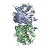 1ns0C 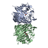 1ns4C 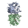 1ns7C  1ns8C 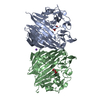 1nsmC 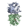 1nsrC  1nssC  1nsuC  1nsvC  1nsxC 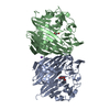 1nszC  1l7jS C: citing same article ( S: Starting model for refinement |
|---|---|
| Similar structure data |


-Links
 PDBj
PDBj


- Assembly
| Deposited unit | 
| ||||||||
|---|---|---|---|---|---|---|---|---|---|
| 1 |
| ||||||||
| Unit cell |
|
-Components
| #1: Protein | Mass: 38564.922 Da / Num. of mol.: 2 / Mutation: E304A Source method: isolated from a genetically manipulated source Source: (gene. exp.) Lactococcus lactis (lactic acid bacteria)Plasmid: PET28 / Production host: #2: Sugar |   Type: D-saccharide, beta linking / Mass: 180.156 Da / Num. of mol.: 2 Type: D-saccharide, beta linking / Mass: 180.156 Da / Num. of mol.: 2Source method: isolated from a genetically manipulated source Formula: C6H12O6 #3: Chemical | ChemComp-NI / |   Mass: 58.693 Da / Num. of mol.: 1 / Source method: obtained synthetically / Formula: Ni Mass: 58.693 Da / Num. of mol.: 1 / Source method: obtained synthetically / Formula: Ni#4: Water | ChemComp-HOH / |  Mass: 18.015 Da / Num. of mol.: 402 / Source method: isolated from a natural source / Formula: H2O Mass: 18.015 Da / Num. of mol.: 402 / Source method: isolated from a natural source / Formula: H2O |
|---|
-Experimental details
-Experiment
| Experiment | Method: X-RAY DIFFRACTION / Number of used crystals: 1 |
|---|
- Sample preparation
| Crystal | Density Matthews: 2.23 Å3/Da / Density % sol: 44.48 % | ||||||||||||||||||||||||
|---|---|---|---|---|---|---|---|---|---|---|---|---|---|---|---|---|---|---|---|---|---|---|---|---|---|
| Crystal grow | Temperature: 277 K / Method: vapor diffusion, hanging drop / pH: 6 Details: MES, methylether PEG-5000, NaCl, pH 6.00, VAPOR DIFFUSION, HANGING DROP, temperature 277K | ||||||||||||||||||||||||
| Crystal grow | *PLUS Temperature: 4 ℃ / pH: 6 / Method: vapor diffusion | ||||||||||||||||||||||||
| Components of the solutions | *PLUS
|
-Data collection
| Diffraction | Mean temperature: 277 K |
|---|---|
| Diffraction source | Source: ROTATING ANODE / Type: RIGAKU RU200 / Wavelength: 1.54178 Å |
| Detector | Type: SIEMENS HI-STAR / Detector: AREA DETECTOR / Date: Mar 25, 2002 / Details: GOEBEL MIRRORS |
| Radiation | Monochromator: GOEBEL OPTICS / Protocol: SINGLE WAVELENGTH / Monochromatic (M) / Laue (L): M / Scattering type: x-ray |
| Radiation wavelength | Wavelength: 1.54178 Å / Relative weight: 1 |
| Reflection | Resolution: 1.95→30 Å / Num. all: 50827 / Num. obs: 50827 / % possible obs: 94.1 % / Observed criterion σ(F): 0 / Observed criterion σ(I): 0 / Redundancy: 2.5 % / Rsym value: 0.0702 / Net I/σ(I): 10.5 |
| Reflection shell | Resolution: 1.95→2.04 Å / Redundancy: 1.9 % / Mean I/σ(I) obs: 2.9 / Num. unique all: 5840 / Rsym value: 0.248 / % possible all: 86.8 |
| Reflection | *PLUS Highest resolution: 1.95 Å / Lowest resolution: 30 Å / Rmerge(I) obs: 0.0702 |
- Processing
| Software |
| |||||||||||||||||||||||||
|---|---|---|---|---|---|---|---|---|---|---|---|---|---|---|---|---|---|---|---|---|---|---|---|---|---|---|
| Refinement | Method to determine structure: FOURIER SYNTHESIS Starting model: PDB ENTRY 1L7J Resolution: 1.95→30 Å / σ(F): 0 / σ(I): 0 / Stereochemistry target values: ENGH & HUBER
| |||||||||||||||||||||||||
| Refinement step | Cycle: LAST / Resolution: 1.95→30 Å
| |||||||||||||||||||||||||
| Refine LS restraints |
| |||||||||||||||||||||||||
| Refinement | *PLUS Lowest resolution: 30 Å / Num. reflection obs: 45793 | |||||||||||||||||||||||||
| Solvent computation | *PLUS | |||||||||||||||||||||||||
| Displacement parameters | *PLUS | |||||||||||||||||||||||||
| Refine LS restraints | *PLUS
|
