 Movie
Movie Controller

[English] 日本語
 Yorodumi
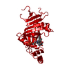
- PDB-1nrg: Structure and Properties of Recombinant Human Pyridoxine-5'-Phosp... -
+ Open data

- Basic information
| Entry | Database: PDB / ID: 1nrg | ||||||
|---|---|---|---|---|---|---|---|
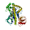
| Title | Structure and Properties of Recombinant Human Pyridoxine-5'-Phosphate Oxidase | ||||||
 Components Components | pyridoxine 5'-phosphate oxidase | ||||||
 Keywords Keywords | OXIDOREDUCTASE / PLP / FMN / pyridoxine-5'-phosphate / oxidase | ||||||
| Function / homology |  Function and homology information Function and homology informationpyridoxamine metabolic process / pyridoxal 5'-phosphate synthase / Vitamin B6 activation to pyridoxal phosphate / pyridoxamine phosphate oxidase activity / pyridoxal 5'-phosphate biosynthetic process / pyridoxine biosynthetic process / FMN binding / pyridoxal phosphate binding / protein homodimerization activity / mitochondrion / cytosol Similarity search - Function | ||||||
| Biological species |  Homo sapiens (human) Homo sapiens (human) | ||||||
| Method |  X-RAY DIFFRACTION / MOLECULAR REPLACEMENT / Resolution: 1.95 Å X-RAY DIFFRACTION / MOLECULAR REPLACEMENT / Resolution: 1.95 Å | ||||||
 Authors Authors | Musayev, F.N. / di Salvo, M.L. / Ko, T.P. / Schirch, V. / Safo, M.K. | ||||||
 Citation Citation | Journal: Protein Sci. / Year: 2003 Title: Structure and properties of recombinant human pyridoxine 5'-phosphate oxidase. Authors: Musayev, F.N. / Di Salvo, M.L. / Ko, T.P. / Schirch, V. / Safo, M.K. | ||||||
| History |
|
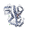
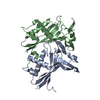
- Structure visualization
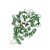
| Structure viewer | Molecule: MolmilJmol/JSmol |
|---|
- Downloads & links
-Download
| PDBx/mmCIF format | 1nrg.cif.gz | 66.2 KB | Display | PDBx/mmCIF format |
|---|---|---|---|---|
| PDB format | pdb1nrg.ent.gz | 46.9 KB | Display | PDB format |
| PDBx/mmJSON format | 1nrg.json.gz | Tree view | PDBx/mmJSON format | |
| Others |  Other downloads Other downloads |
-Validation report
| Arichive directory | https://data.pdbj.org/pub/pdb/validation_reports/nr/1nrgftp://data.pdbj.org/pub/pdb/validation_reports/nr/1nrg | HTTPS FTP |
|---|
-Related structure data
| Related structure data |  1g79S S: Starting model for refinement |
|---|---|
| Similar structure data |


-Links
 PDBj
PDBj


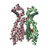
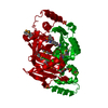
- Assembly
| Deposited unit | 
| ||||||||
|---|---|---|---|---|---|---|---|---|---|
| 1 | 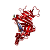
| ||||||||
| Unit cell |
| ||||||||
| Details | The biological assembly is a dimer, which can be constructed from chain A and its symmetry-related partner generated by X-Y, -Y, 2/3-Z |
-Components
-Protein , 1 types, 1 molecules A
| #1: Protein | Mass: 30029.025 Da / Num. of mol.: 1 Source method: isolated from a genetically manipulated source Source: (gene. exp.) Homo sapiens (human) / Gene: Human / Plasmid: pET28 / Production host:  |
|---|
-Non-polymers , 5 types, 200 molecules 


| #2: Chemical | ChemComp-PO4 /  Mass: 94.971 Da / Num. of mol.: 1 / Source method: obtained synthetically / Formula: PO4 Mass: 94.971 Da / Num. of mol.: 1 / Source method: obtained synthetically / Formula: PO4 |
|---|---|
| #3: Chemical | ChemComp-FMN /  Mass: 456.344 Da / Num. of mol.: 1 / Source method: obtained synthetically / Formula: C17H21N4O9P Mass: 456.344 Da / Num. of mol.: 1 / Source method: obtained synthetically / Formula: C17H21N4O9P |
| #4: Chemical | ChemComp-PLP /  Mass: 247.142 Da / Num. of mol.: 1 / Source method: obtained synthetically / Formula: C8H10NO6P Mass: 247.142 Da / Num. of mol.: 1 / Source method: obtained synthetically / Formula: C8H10NO6P |
| #5: Chemical | ChemComp-BME /  Mass: 78.133 Da / Num. of mol.: 1 / Source method: obtained synthetically / Formula: C2H6OS Mass: 78.133 Da / Num. of mol.: 1 / Source method: obtained synthetically / Formula: C2H6OS |
| #6: Water | ChemComp-HOH / Mass: 18.015 Da / Num. of mol.: 196 / Source method: isolated from a natural source / Formula: H2O |
-Experimental details
-Experiment
| Experiment | Method: X-RAY DIFFRACTION / Number of used crystals: 1 |
|---|
- Sample preparation
| Crystal | Density Matthews: 2.1 Å3/Da / Density % sol: 40.86 % | ||||||||||||||||||||||||||||||||||||||||||
|---|---|---|---|---|---|---|---|---|---|---|---|---|---|---|---|---|---|---|---|---|---|---|---|---|---|---|---|---|---|---|---|---|---|---|---|---|---|---|---|---|---|---|---|
| Crystal grow | Temperature: 298 K / Method: vapor diffusion, hanging drop / pH: 7.5 Details: potassium/sodium tartarate, potassium phosphate, 2-mercaptoethanol, pH 7.5, VAPOR DIFFUSION, HANGING DROP, temperature 298K | ||||||||||||||||||||||||||||||||||||||||||
| Crystal grow | *PLUS | ||||||||||||||||||||||||||||||||||||||||||
| Components of the solutions | *PLUS
|
-Data collection
| Diffraction | Mean temperature: 100 K |
|---|---|
| Diffraction source | Source: ROTATING ANODE / Type: RIGAKU RU200 / Wavelength: 1.5418 Å |
| Detector | Type: RIGAKU RAXIS II / Detector: IMAGE PLATE / Date: Oct 16, 2002 / Details: mirrors |
| Radiation | Monochromator: MSC Confocal Mirrors / Protocol: SINGLE WAVELENGTH / Monochromatic (M) / Laue (L): M / Scattering type: x-ray |
| Radiation wavelength | Wavelength: 1.5418 Å / Relative weight: 1 |
| Reflection | Resolution: 1.95→71.7 Å / Num. all: 17885 / Num. obs: 16966 / % possible obs: 94.9 % / Observed criterion σ(F): 0 / Observed criterion σ(I): 0 / Redundancy: 5.59 % / Biso Wilson estimate: 23.4 Å2 / Rmerge(I) obs: 0.0654 / Net I/σ(I): 14.7 |
| Reflection shell | Resolution: 1.95→2 Å / Redundancy: 3.87 % / Rmerge(I) obs: 0.366 / Mean I/σ(I) obs: 1.63 / Num. unique all: 1226 / % possible all: 95.8 |
| Reflection | *PLUS Lowest resolution: 71 Å / Num. measured all: 94811 / Rmerge(I) obs: 0.065 |
| Reflection shell | *PLUS % possible obs: 95.8 % / Num. unique obs: 1226 / Num. measured obs: 4746 |
- Processing
| Software |
| ||||||||||||||||||||||||||||||||||||
|---|---|---|---|---|---|---|---|---|---|---|---|---|---|---|---|---|---|---|---|---|---|---|---|---|---|---|---|---|---|---|---|---|---|---|---|---|---|
| Refinement | Method to determine structure: MOLECULAR REPLACEMENT Starting model: E. coli PNPOx structure, PDB code 1G79 Resolution: 1.95→45.7 Å / Rfactor Rfree error: 0.009 / Isotropic thermal model: RESTRAINED / Cross valid method: THROUGHOUT / σ(F): 0 / Stereochemistry target values: Engh & Huber
| ||||||||||||||||||||||||||||||||||||
| Solvent computation | Solvent model: FLAT MODEL / Bsol: 66.1042 Å2 / ksol: 0.35217 e/Å3 | ||||||||||||||||||||||||||||||||||||
| Displacement parameters | Biso mean: 44.3 Å2
| ||||||||||||||||||||||||||||||||||||
| Refine analyze |
| ||||||||||||||||||||||||||||||||||||
| Refinement step | Cycle: LAST / Resolution: 1.95→45.7 Å
| ||||||||||||||||||||||||||||||||||||
| Refine LS restraints |
| ||||||||||||||||||||||||||||||||||||
| LS refinement shell | Resolution: 1.95→2.07 Å / Rfactor Rfree error: 0.027 / Total num. of bins used: 6
| ||||||||||||||||||||||||||||||||||||
| Xplor file |
| ||||||||||||||||||||||||||||||||||||
| Refinement | *PLUS Lowest resolution: 45.7 Å / % reflection Rfree: 5 % / Rfactor Rfree: 0.252 | ||||||||||||||||||||||||||||||||||||
| Solvent computation | *PLUS | ||||||||||||||||||||||||||||||||||||
| Displacement parameters | *PLUS | ||||||||||||||||||||||||||||||||||||
| Refine LS restraints | *PLUS
| ||||||||||||||||||||||||||||||||||||
| LS refinement shell | *PLUS Lowest resolution: 1.99 Å / Rfactor Rfree: 0.321 / Rfactor Rwork: 0.311 / Num. reflection obs: 1094 |
