 Movie
Movie Controller

[English] 日本語
 Yorodumi
- PDB-1npm: NEUROPSIN, A SERINE PROTEASE EXPRESSED IN THE LIMBIC SYSTEM OF MO... -
+ Open data

- Basic information
| Entry | Database: PDB / ID: 1npm | ||||||
|---|---|---|---|---|---|---|---|
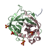
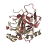
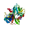
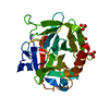
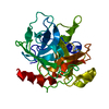
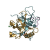
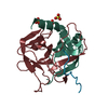
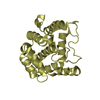
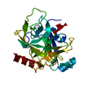
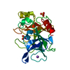
| Title | NEUROPSIN, A SERINE PROTEASE EXPRESSED IN THE LIMBIC SYSTEM OF MOUSE BRAIN | ||||||
 Components Components | NEUROPSIN | ||||||
 Keywords Keywords | SERINE PROTEINASE / GLYCOPROTEIN | ||||||
| Function / homology |  Function and homology information Function and homology informationFormation of the cornified envelope / kallikrein 8 / regulation of synapse organization / keratinocyte proliferation / serine protease inhibitor complex / neuron projection morphogenesis / synapse organization / response to wounding / memory / peptidase activity ...Formation of the cornified envelope / kallikrein 8 / regulation of synapse organization / keratinocyte proliferation / serine protease inhibitor complex / neuron projection morphogenesis / synapse organization / response to wounding / memory / peptidase activity / serine-type endopeptidase activity / proteolysis / extracellular space / cytoplasm Similarity search - Function | ||||||
| Biological species |  | ||||||
| Method |  X-RAY DIFFRACTION / SYNCHROTRON / MOLECULAR REPLACEMENT / Resolution: 2.1 Å X-RAY DIFFRACTION / SYNCHROTRON / MOLECULAR REPLACEMENT / Resolution: 2.1 Å | ||||||
 Authors Authors | Kishi, T. / Kato, M. / Shimizu, T. / Kato, K. / Matsumoto, K. / Yoshida, S. / Shiosaka, S. / Hakoshima, T. | ||||||
 Citation Citation | Journal: J.Biol.Chem. / Year: 1999 Title: Crystal structure of neuropsin, a hippocampal protease involved in kindling epileptogenesis. Authors: Kishi, T. / Kato, M. / Shimizu, T. / Kato, K. / Matsumoto, K. / Yoshida, S. / Shiosaka, S. / Hakoshima, T. #1: Journal: J.Struct.Biol. / Year: 1997Title: Crystallization and Preliminary X-Ray Analysis of Neuropsin, a Serine Protease Expressed in the Limbic System of Mouse Brain Authors: Kishi, T. / Kato, M. / Shimizu, T. / Kato, K. / Matsumoto, K. / Yoshida, S. / Shiosaka, S. / Hakoshima, T. | ||||||
| History |
|
- Structure visualization
| Structure viewer | Molecule: MolmilJmol/JSmol |
|---|
- Downloads & links
-Download
| PDBx/mmCIF format | 1npm.cif.gz | 101.4 KB | Display | PDBx/mmCIF format |
|---|---|---|---|---|
| PDB format | pdb1npm.ent.gz | 76.7 KB | Display | PDB format |
| PDBx/mmJSON format | 1npm.json.gz | Tree view | PDBx/mmJSON format | |
| Others |  Other downloads Other downloads |
-Validation report
| Summary document | 1npm_validation.pdf.gz | 393.2 KB | Display | wwPDB validaton report |
|---|---|---|---|---|
| Full document | 1npm_full_validation.pdf.gz | 400.8 KB | Display | |
| Data in XML | 1npm_validation.xml.gz | 10.9 KB | Display | |
| Data in CIF | 1npm_validation.cif.gz | 16.7 KB | Display | |
| Arichive directory | https://data.pdbj.org/pub/pdb/validation_reports/np/1npmftp://data.pdbj.org/pub/pdb/validation_reports/np/1npm | HTTPS FTP |
-Related structure data
| Related structure data |  4ptp S: Starting model for refinement |
|---|---|
| Similar structure data |
-Links
 PDBj
PDBj
- Assembly
| Deposited unit | 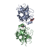
| ||||||||
|---|---|---|---|---|---|---|---|---|---|
| 1 | 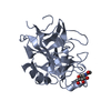
| ||||||||
| 2 | 
| ||||||||
| Unit cell |
| ||||||||
| Noncrystallographic symmetry (NCS) | NCS oper: (Code: given Matrix: (1, -0.00042, 0.00043), Vector: |
-Components
| #1: Protein | Mass: 24704.045 Da / Num. of mol.: 2 Source method: isolated from a genetically manipulated source Source: (gene. exp.)   Spodoptera frugiperda (fall armyworm) / References: UniProt: Q61955 Spodoptera frugiperda (fall armyworm) / References: UniProt: Q61955#2: Sugar |   Type: D-saccharide, beta linking / Mass: 221.208 Da / Num. of mol.: 2 Type: D-saccharide, beta linking / Mass: 221.208 Da / Num. of mol.: 2Source method: isolated from a genetically manipulated source Formula: C8H15NO6 #3: Water | ChemComp-HOH / |  Mass: 18.015 Da / Num. of mol.: 194 / Source method: isolated from a natural source / Formula: H2O Mass: 18.015 Da / Num. of mol.: 194 / Source method: isolated from a natural source / Formula: H2OHas protein modification | Y | |
|---|
-Experimental details
-Experiment
| Experiment | Method: X-RAY DIFFRACTION / Number of used crystals: 2 |
|---|
- Sample preparation
| Crystal | Density Matthews: 2.5 Å3/Da / Density % sol: 51 % | ||||||||||||||||||||||||
|---|---|---|---|---|---|---|---|---|---|---|---|---|---|---|---|---|---|---|---|---|---|---|---|---|---|
| Crystal grow | pH: 7 / Details: pH 7.0 | ||||||||||||||||||||||||
| Crystal grow | *PLUS Temperature: 4 ℃ / Method: vapor diffusion, hanging drop | ||||||||||||||||||||||||
| Components of the solutions | *PLUS
|
-Data collection
| Diffraction | Mean temperature: 277 K |
|---|---|
| Diffraction source | Source: SYNCHROTRON / Site: Photon Factory  / Beamline: BL-18B / Wavelength: 1 / Beamline: BL-18B / Wavelength: 1 |
| Detector | Detector: IMAGE PLATE / Date: Nov 1, 1996 |
| Radiation | Monochromatic (M) / Laue (L): M / Scattering type: x-ray |
| Radiation wavelength | Wavelength: 1 Å / Relative weight: 1 |
| Reflection | Highest resolution: 2.1 Å / Num. obs: 24944 / % possible obs: 90 % / Observed criterion σ(I): 1 / Redundancy: 2.5 % / Biso Wilson estimate: 30 Å2 / Rmerge(I) obs: 0.06 / Net I/σ(I): 8.5 |
| Reflection shell | Resolution: 2.1→2.24 Å / Redundancy: 1.8 % / Rmerge(I) obs: 0.198 / Mean I/σ(I) obs: 3.1 / % possible all: 74.5 |
| Reflection | *PLUS % possible obs: 90.7 % |
| Reflection shell | *PLUS % possible obs: 74.5 % |
- Processing
| Software |
| ||||||||||||||||||||||||||||||||||||||||||||||||||||||||||||
|---|---|---|---|---|---|---|---|---|---|---|---|---|---|---|---|---|---|---|---|---|---|---|---|---|---|---|---|---|---|---|---|---|---|---|---|---|---|---|---|---|---|---|---|---|---|---|---|---|---|---|---|---|---|---|---|---|---|---|---|---|---|
| Refinement | Method to determine structure: MOLECULAR REPLACEMENT Starting model: PDB ENTRY 4PTP 4ptp Resolution: 2.1→100 Å / Data cutoff high absF: 10000000 / Data cutoff low absF: 0.001 / Cross valid method: THROUGHOUT / σ(F): 1
| ||||||||||||||||||||||||||||||||||||||||||||||||||||||||||||
| Displacement parameters | Biso mean: 31.1 Å2 | ||||||||||||||||||||||||||||||||||||||||||||||||||||||||||||
| Refine analyze | Luzzati coordinate error obs: 0.34 Å | ||||||||||||||||||||||||||||||||||||||||||||||||||||||||||||
| Refinement step | Cycle: LAST / Resolution: 2.1→100 Å
| ||||||||||||||||||||||||||||||||||||||||||||||||||||||||||||
| Refine LS restraints |
| ||||||||||||||||||||||||||||||||||||||||||||||||||||||||||||
| LS refinement shell | Resolution: 2.1→2.2 Å / Total num. of bins used: 8
| ||||||||||||||||||||||||||||||||||||||||||||||||||||||||||||
| Xplor file |
| ||||||||||||||||||||||||||||||||||||||||||||||||||||||||||||
| Software | *PLUS Name: X-PLOR / Version: 3.851 / Classification: refinement | ||||||||||||||||||||||||||||||||||||||||||||||||||||||||||||
| Refinement | *PLUS | ||||||||||||||||||||||||||||||||||||||||||||||||||||||||||||
| Solvent computation | *PLUS | ||||||||||||||||||||||||||||||||||||||||||||||||||||||||||||
| Displacement parameters | *PLUS Biso mean: 30.8 Å2 | ||||||||||||||||||||||||||||||||||||||||||||||||||||||||||||
| Refine LS restraints | *PLUS
| ||||||||||||||||||||||||||||||||||||||||||||||||||||||||||||
| LS refinement shell | *PLUS Rfactor obs: 0.281 |
