 Movie
Movie Controller

[English] 日本語
 Yorodumi
- PDB-3mu1: Comparison of the character and the speed of X-ray-induced struct... -
+ Open data

- Basic information
| Entry | Database: PDB / ID: 3mu1 | ||||||
|---|---|---|---|---|---|---|---|
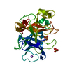
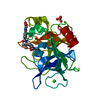
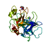
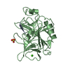
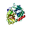
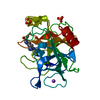
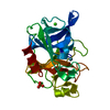
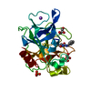
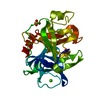
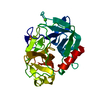
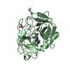
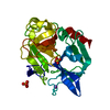
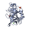
| Title | Comparison of the character and the speed of X-ray-induced structural changes of porcine pancreatic elastase at two temperatures, 100 and 15K. The data set was collected from region A of the crystal. Fifth step of radiation damage | ||||||
 Components Components | Chymotrypsin-like elastase family member 1 | ||||||
 Keywords Keywords | HYDROLASE / radiation damage / disulfide bridge / atomic resolution | ||||||
| Function / homology |  Function and homology information Function and homology informationpancreatic elastase / serine-type endopeptidase activity / proteolysis / : / metal ion binding Similarity search - Function | ||||||
| Biological species |  | ||||||
| Method |  X-RAY DIFFRACTION / SYNCHROTRON / MOLECULAR REPLACEMENT / Resolution: 1.74 Å X-RAY DIFFRACTION / SYNCHROTRON / MOLECULAR REPLACEMENT / Resolution: 1.74 Å | ||||||
 Authors Authors | Petrova, T. / Ginell, S. / Mitschler, A. / Cousido-Siah, A. / Hazemann, I. / Podjarny, A. / Joachimiak, A. | ||||||
 Citation Citation | Journal: Acta Crystallogr.,Sect.D / Year: 2010 Title: X-ray-induced deterioration of disulfide bridges at atomic resolution. Authors: Petrova, T. / Ginell, S. / Mitschler, A. / Kim, Y. / Lunin, V.Y. / Joachimiak, G. / Cousido-Siah, A. / Hazemann, I. / Podjarny, A. / Lazarski, K. / Joachimiak, A. | ||||||
| History |
|
- Structure visualization
| Structure viewer | Molecule: MolmilJmol/JSmol |
|---|
- Downloads & links
-Download
| PDBx/mmCIF format | 3mu1.cif.gz | 77.8 KB | Display | PDBx/mmCIF format |
|---|---|---|---|---|
| PDB format | pdb3mu1.ent.gz | 55.9 KB | Display | PDB format |
| PDBx/mmJSON format | 3mu1.json.gz | Tree view | PDBx/mmJSON format | |
| Others |  Other downloads Other downloads |
-Validation report
| Arichive directory | https://data.pdbj.org/pub/pdb/validation_reports/mu/3mu1ftp://data.pdbj.org/pub/pdb/validation_reports/mu/3mu1 | HTTPS FTP |
|---|
-Related structure data
| Related structure data |  3mnbC  3mncC  3mnsC  3mnxC 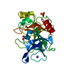 3mo3C 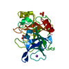 3mo6C  3mo9C 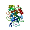 3mocC  3mtyC 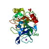 3mu0C 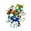 3mu4C 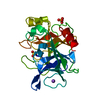 3mu5C  3mu8C  3oddC  3odfC 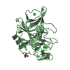 1gvkS  3mt4 S: Starting model for refinement C: citing same article ( |
|---|---|
| Similar structure data |


-Links
 PDBj
PDBj
- Assembly
| Deposited unit | 
| ||||||||
|---|---|---|---|---|---|---|---|---|---|
| 1 |
| ||||||||
| Unit cell |
|
-Components
| #1: Protein | Mass: 25928.031 Da / Num. of mol.: 1 / Source method: isolated from a natural source / Source: (natural) | ||
|---|---|---|---|
| #2: Chemical | ChemComp-NA /   Mass: 22.990 Da / Num. of mol.: 1 / Source method: obtained synthetically / Formula: Na Mass: 22.990 Da / Num. of mol.: 1 / Source method: obtained synthetically / Formula: Na | ||
| #3: Chemical |   Mass: 96.063 Da / Num. of mol.: 2 / Source method: obtained synthetically / Formula: SO4 Mass: 96.063 Da / Num. of mol.: 2 / Source method: obtained synthetically / Formula: SO4#4: Water | ChemComp-HOH / |  Mass: 18.015 Da / Num. of mol.: 479 / Source method: isolated from a natural source / Formula: H2O Mass: 18.015 Da / Num. of mol.: 479 / Source method: isolated from a natural source / Formula: H2O |
-Experimental details
-Experiment
| Experiment | Method: X-RAY DIFFRACTION / Number of used crystals: 1 |
|---|
- Sample preparation
| Crystal | Density Matthews: 2.07 Å3/Da / Density % sol: 40.71 % |
|---|---|
| Crystal grow | Temperature: 291 K / Method: vapor diffusion, sitting drop / pH: 7.5 Details: The initial concentration of the protein was 20 mg/ml in 10% glycerol solution. The reservoir contained a 250 mM Na2SO4. For cryo-protection, it was supplemented with 25% glycerol, pH 7.5, ...Details: The initial concentration of the protein was 20 mg/ml in 10% glycerol solution. The reservoir contained a 250 mM Na2SO4. For cryo-protection, it was supplemented with 25% glycerol, pH 7.5, VAPOR DIFFUSION, SITTING DROP, temperature 291K |
-Data collection
| Diffraction | Mean temperature: 100 K |
|---|---|
| Diffraction source | Source: SYNCHROTRON / Site: APS  / Beamline: 19-ID / Wavelength: 0.97895 Å / Beamline: 19-ID / Wavelength: 0.97895 Å |
| Detector | Type: ADSC QUANTUM 315 / Detector: CCD / Date: Mar 20, 2008 Details: 1.02-M FLAT MIRROR MADE OF ZERODUR PROVIDING VERTICAL FOCUSING AND REJECTION OF HARMONIC CONTAMINATION |
| Radiation | Monochromator: DOUBLE CRYSTAL MONOCHROMATOR UTILIZING A SI-111 AND SAGITAL HORIZONTAL FOCUSING Protocol: SINGLE WAVELENGTH / Monochromatic (M) / Laue (L): M / Scattering type: x-ray |
| Radiation wavelength | Wavelength: 0.97895 Å / Relative weight: 1 |
| Reflection | Resolution: 1.74→50 Å / Num. obs: 22233 / % possible obs: 99.5 % / Observed criterion σ(I): 2 / Redundancy: 4.4 % / Biso Wilson estimate: 20.22 Å2 / Rmerge(I) obs: 0.027 / Net I/σ(I): 30.52 |
| Reflection shell | Resolution: 1.74→1.81 Å / Redundancy: 4.5 % / Rmerge(I) obs: 0.375 / Mean I/σ(I) obs: 3.54 / Num. unique all: 2190 / % possible all: 99.7 |
- Processing
| Software |
| |||||||||||||||||||||||||||||||||||||||||||||||||||||||||||||||
|---|---|---|---|---|---|---|---|---|---|---|---|---|---|---|---|---|---|---|---|---|---|---|---|---|---|---|---|---|---|---|---|---|---|---|---|---|---|---|---|---|---|---|---|---|---|---|---|---|---|---|---|---|---|---|---|---|---|---|---|---|---|---|---|---|
| Refinement | Method to determine structure: MOLECULAR REPLACEMENT Starting model: PDB entry 1GVK Resolution: 1.74→18.773 Å / SU ML: 0.23 / Isotropic thermal model: Isotropic / σ(F): 0.17 / Stereochemistry target values: ML
| |||||||||||||||||||||||||||||||||||||||||||||||||||||||||||||||
| Solvent computation | Shrinkage radii: 0.9 Å / VDW probe radii: 1.11 Å / Solvent model: FLAT BULK SOLVENT MODEL / Bsol: 126.469 Å2 / ksol: 0.402 e/Å3 | |||||||||||||||||||||||||||||||||||||||||||||||||||||||||||||||
| Displacement parameters | Biso mean: 25.02 Å2
| |||||||||||||||||||||||||||||||||||||||||||||||||||||||||||||||
| Refine analyze | Luzzati coordinate error obs: 0.23 Å | |||||||||||||||||||||||||||||||||||||||||||||||||||||||||||||||
| Refinement step | Cycle: LAST / Resolution: 1.74→18.773 Å
| |||||||||||||||||||||||||||||||||||||||||||||||||||||||||||||||
| Refine LS restraints |
| |||||||||||||||||||||||||||||||||||||||||||||||||||||||||||||||
| LS refinement shell | Refine-ID: X-RAY DIFFRACTION
|
