 Movie
Movie Controller

[English] 日本語
 Yorodumi
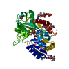
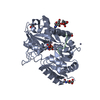
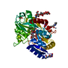
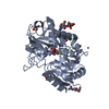
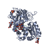
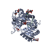
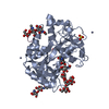
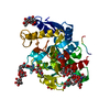
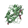
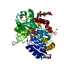
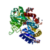
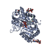
- PDB-1nkx: CRYSTAL STRUCTURE OF A PROTEOLYTICALLY GENERATED FUNCTIONAL MONOF... -
+ Open data

- Basic information
| Entry | Database: PDB / ID: 1nkx | |||||||||
|---|---|---|---|---|---|---|---|---|---|---|
| Title | CRYSTAL STRUCTURE OF A PROTEOLYTICALLY GENERATED FUNCTIONAL MONOFERRIC C-LOBE OF BOVINE LACTOFERRIN AT 1.9A RESOLUTION | |||||||||
 Components Components | LACTOTRANSFERRIN | |||||||||
 Keywords Keywords | TRANSPORT PROTEIN / lactoferrin / C-lobe / fragment / trypsin | |||||||||
| Function / homology |  Function and homology information Function and homology informationMetal sequestration by antimicrobial proteins / Antimicrobial peptides / negative regulation of tumor necrosis factor (ligand) superfamily member 11 production / negative regulation of single-species biofilm formation in or on host organism / positive regulation of bone mineralization involved in bone maturation / negative regulation of osteoclast development / specific granule / negative regulation of lipopolysaccharide-mediated signaling pathway / positive regulation of chondrocyte proliferation / antifungal humoral response ...Metal sequestration by antimicrobial proteins / Antimicrobial peptides / negative regulation of tumor necrosis factor (ligand) superfamily member 11 production / negative regulation of single-species biofilm formation in or on host organism / positive regulation of bone mineralization involved in bone maturation / negative regulation of osteoclast development / specific granule / negative regulation of lipopolysaccharide-mediated signaling pathway / positive regulation of chondrocyte proliferation / antifungal humoral response / regulation of tumor necrosis factor production / bone morphogenesis / Neutrophil degranulation / positive regulation of osteoblast proliferation / Hydrolases; Acting on peptide bonds (peptidases); Serine endopeptidases / positive regulation of osteoblast differentiation / cysteine-type endopeptidase inhibitor activity / regulation of cytokine production / ossification / innate immune response in mucosa / iron ion transport / recycling endosome / antibacterial humoral response / early endosome / iron ion binding / serine-type endopeptidase activity / signaling receptor binding / negative regulation of apoptotic process / proteolysis / : / plasma membrane Similarity search - Function | |||||||||
| Biological species |  | |||||||||
| Method |  X-RAY DIFFRACTION / MOLECULAR REPLACEMENT / Resolution: 1.9 Å X-RAY DIFFRACTION / MOLECULAR REPLACEMENT / Resolution: 1.9 Å | |||||||||
 Authors Authors | Sharma, S. / Jasti, J. / Kumar, J. / Mohanty, A.K. / Singh, T.P. | |||||||||
 Citation Citation | Journal: J.Mol.Biol. / Year: 2003 Title: Crystal Structure of a Proteolytically Generated Functional Monoferric C-lobe of Bovine Lactoferrin at 1.9A Resolution Authors: Sharma, S. / Jasti, J. / Kumar, J. / Mohanty, A.K. / Singh, T.P. | |||||||||
| History |
|
- Structure visualization
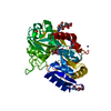
| Structure viewer | Molecule: MolmilJmol/JSmol |
|---|
- Downloads & links
-Download
| PDBx/mmCIF format | 1nkx.cif.gz | 90.1 KB | Display | PDBx/mmCIF format |
|---|---|---|---|---|
| PDB format | pdb1nkx.ent.gz | 65.4 KB | Display | PDB format |
| PDBx/mmJSON format | 1nkx.json.gz | Tree view | PDBx/mmJSON format | |
| Others |  Other downloads Other downloads |
-Validation report
| Arichive directory | https://data.pdbj.org/pub/pdb/validation_reports/nk/1nkxftp://data.pdbj.org/pub/pdb/validation_reports/nk/1nkx | HTTPS FTP |
|---|
-Related structure data
| Related structure data | 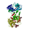 1blfS S: Starting model for refinement |
|---|---|
| Similar structure data |


-Links
 PDBj
PDBj


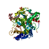
- Assembly
| Deposited unit | 
| ||||||||
|---|---|---|---|---|---|---|---|---|---|
| 1 |
| ||||||||
| Unit cell |
|
-Components
-Protein , 1 types, 1 molecules A
| #1: Protein | Mass: 38026.961 Da / Num. of mol.: 1 / Source method: isolated from a natural source / Details: milk / Source: (natural) |
|---|
-Sugars , 3 types, 3 molecules 
| #2: Polysaccharide | beta-D-mannopyranose-(1-4)-2-acetamido-2-deoxy-beta-D-glucopyranose-(1-4)-2-acetamido-2-deoxy-beta- ...beta-D-mannopyranose-(1-4)-2-acetamido-2-deoxy-beta-D-glucopyranose-(1-4)-2-acetamido-2-deoxy-beta-D-glucopyranose Source method: isolated from a genetically manipulated source |
|---|---|
| #3: Polysaccharide | alpha-D-mannopyranose-(1-4)-alpha-D-mannopyranose-(1-4)-alpha-D-mannopyranose-(1-4)-beta-D- ...alpha-D-mannopyranose-(1-4)-alpha-D-mannopyranose-(1-4)-alpha-D-mannopyranose-(1-4)-beta-D-mannopyranose-(1-4)-2-acetamido-2-deoxy-beta-D-glucopyranose-(1-4)-2-acetamido-2-deoxy-beta-D-glucopyranose Source method: isolated from a genetically manipulated source |
| #4: Sugar | ChemComp-NAG /  Type: D-saccharide, beta linking / Mass: 221.208 Da / Num. of mol.: 1 Type: D-saccharide, beta linking / Mass: 221.208 Da / Num. of mol.: 1Source method: isolated from a genetically manipulated source Formula: C8H15NO6 |
-Non-polymers , 4 types, 230 molecules 


| #5: Chemical | ChemComp-FE /  Mass: 55.845 Da / Num. of mol.: 1 / Source method: obtained synthetically / Formula: Fe Mass: 55.845 Da / Num. of mol.: 1 / Source method: obtained synthetically / Formula: Fe | ||||
|---|---|---|---|---|---|
| #6: Chemical |  Mass: 65.409 Da / Num. of mol.: 2 / Source method: obtained synthetically / Formula: Zn Mass: 65.409 Da / Num. of mol.: 2 / Source method: obtained synthetically / Formula: Zn#7: Chemical | ChemComp-CO3 / |  Mass: 60.009 Da / Num. of mol.: 1 / Source method: obtained synthetically / Formula: CO3 Mass: 60.009 Da / Num. of mol.: 1 / Source method: obtained synthetically / Formula: CO3#8: Water | ChemComp-HOH / | Mass: 18.015 Da / Num. of mol.: 226 / Source method: isolated from a natural source / Formula: H2O |
-Details
| Has protein modification | Y |
|---|
-Experimental details
-Experiment
| Experiment | Method: X-RAY DIFFRACTION / Number of used crystals: 1 |
|---|
- Sample preparation
| Crystal | Density Matthews: 2.64 Å3/Da / Density % sol: 53.41 % | ||||||||||||||||||||||||||||||
|---|---|---|---|---|---|---|---|---|---|---|---|---|---|---|---|---|---|---|---|---|---|---|---|---|---|---|---|---|---|---|---|
| Crystal grow | Temperature: 298 K / Method: vapor diffusion / pH: 6.5 Details: 0.1M MES, 25% POLYETHYLENE GLYCOL MONOMETHYL ETHER 550 AND 0.01 M ZINC SULPHATE, pH 6.5, VAPOR DIFFUSION, temperature 298K | ||||||||||||||||||||||||||||||
| Crystal grow | *PLUS Temperature: 25 ℃ / Method: vapor diffusion | ||||||||||||||||||||||||||||||
| Components of the solutions | *PLUS
|
-Data collection
| Diffraction | Mean temperature: 283 K |
|---|---|
| Diffraction source | Source: ROTATING ANODE / Type: RIGAKU RU300 / Wavelength: 1.5418 Å |
| Detector | Type: MARRESEARCH / Detector: IMAGE PLATE / Date: Jul 12, 2002 / Details: mirrors |
| Radiation | Monochromator: YALE MIRRORS / Protocol: SINGLE WAVELENGTH / Monochromatic (M) / Laue (L): M / Scattering type: x-ray |
| Radiation wavelength | Wavelength: 1.5418 Å / Relative weight: 1 |
| Reflection | Resolution: 1.9→20 Å / Num. obs: 29084 / % possible obs: 97 % / Observed criterion σ(F): -3 / Observed criterion σ(I): -3 / Redundancy: 6 % / Biso Wilson estimate: 25.5 Å2 / Rmerge(I) obs: 0.057 / Rsym value: 0.057 / Net I/σ(I): 17.5 |
| Reflection shell | Resolution: 1.9→2 Å / Redundancy: 4 % / Rmerge(I) obs: 0.23 / Mean I/σ(I) obs: 3.6 / Num. unique all: 2482 / Rsym value: 0.23 / % possible all: 84 |
| Reflection | *PLUS Highest resolution: 1.9 Å / Lowest resolution: 20 Å / Redundancy: 20 % / Num. measured all: 172337 |
| Reflection shell | *PLUS Highest resolution: 1.9 Å / Lowest resolution: 2 Å / % possible obs: 84 % / Rmerge(I) obs: 0.23 |
- Processing
| Software |
| ||||||||||||||||||||||||||||||||||||||||||||||||||||||||||||||||||||||||||||||||||||||||||||||||||||||||||||
|---|---|---|---|---|---|---|---|---|---|---|---|---|---|---|---|---|---|---|---|---|---|---|---|---|---|---|---|---|---|---|---|---|---|---|---|---|---|---|---|---|---|---|---|---|---|---|---|---|---|---|---|---|---|---|---|---|---|---|---|---|---|---|---|---|---|---|---|---|---|---|---|---|---|---|---|---|---|---|---|---|---|---|---|---|---|---|---|---|---|---|---|---|---|---|---|---|---|---|---|---|---|---|---|---|---|---|---|---|---|
| Refinement | Method to determine structure: MOLECULAR REPLACEMENT Starting model: PDB ENTRY 1BLF Resolution: 1.9→20 Å / Data cutoff high absF: 730682.69 / Data cutoff low absF: 0 / Isotropic thermal model: Isotropic / Cross valid method: THROUGHOUT / σ(F): 0 / σ(I): 0 / Stereochemistry target values: MAXIMUM LIKELIHOOD / Details: Used weighted full matrix least squares procedure.
| ||||||||||||||||||||||||||||||||||||||||||||||||||||||||||||||||||||||||||||||||||||||||||||||||||||||||||||
| Solvent computation | Bsol: 69.1658 Å2 / ksol: 0.373797 e/Å3 | ||||||||||||||||||||||||||||||||||||||||||||||||||||||||||||||||||||||||||||||||||||||||||||||||||||||||||||
| Displacement parameters | Biso mean: 34.4 Å2
| ||||||||||||||||||||||||||||||||||||||||||||||||||||||||||||||||||||||||||||||||||||||||||||||||||||||||||||
| Refine analyze |
| ||||||||||||||||||||||||||||||||||||||||||||||||||||||||||||||||||||||||||||||||||||||||||||||||||||||||||||
| Refinement step | Cycle: LAST / Resolution: 1.9→20 Å
| ||||||||||||||||||||||||||||||||||||||||||||||||||||||||||||||||||||||||||||||||||||||||||||||||||||||||||||
| Refine LS restraints |
| ||||||||||||||||||||||||||||||||||||||||||||||||||||||||||||||||||||||||||||||||||||||||||||||||||||||||||||
| LS refinement shell | Resolution: 1.9→2 Å / Rfactor Rfree error: 0.021
| ||||||||||||||||||||||||||||||||||||||||||||||||||||||||||||||||||||||||||||||||||||||||||||||||||||||||||||
| Refinement | *PLUS Highest resolution: 1.9 Å / Lowest resolution: 20 Å / Num. reflection obs: 29084 / Rfactor Rfree: 0.221 / Rfactor Rwork: 0.193 | ||||||||||||||||||||||||||||||||||||||||||||||||||||||||||||||||||||||||||||||||||||||||||||||||||||||||||||
| Solvent computation | *PLUS | ||||||||||||||||||||||||||||||||||||||||||||||||||||||||||||||||||||||||||||||||||||||||||||||||||||||||||||
| Displacement parameters | *PLUS | ||||||||||||||||||||||||||||||||||||||||||||||||||||||||||||||||||||||||||||||||||||||||||||||||||||||||||||
| Refine LS restraints | *PLUS
|
