 Movie
Movie Controller

[English] 日本語
 Yorodumi
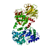
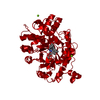
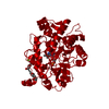
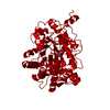
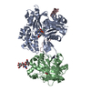
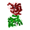
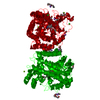
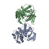
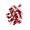
- PDB-1blf: STRUCTURE OF DIFERRIC BOVINE LACTOFERRIN AT 2.8 ANGSTROMS RESOLUTION -
+ Open data

- Basic information
| Entry | Database: PDB / ID: 1blf | |||||||||
|---|---|---|---|---|---|---|---|---|---|---|
| Title | STRUCTURE OF DIFERRIC BOVINE LACTOFERRIN AT 2.8 ANGSTROMS RESOLUTION | |||||||||
 Components Components | LACTOFERRIN | |||||||||
 Keywords Keywords | IRON-BINDING PROTEIN / LACTOFERRIN / TRANSFERRIN / CARBOHYDRATE STRUCTURE | |||||||||
| Function / homology |  Function and homology information Function and homology informationMetal sequestration by antimicrobial proteins / Antimicrobial peptides / negative regulation of tumor necrosis factor (ligand) superfamily member 11 production / negative regulation of single-species biofilm formation in or on host organism / positive regulation of bone mineralization involved in bone maturation / negative regulation of osteoclast development / specific granule / positive regulation of chondrocyte proliferation / antifungal humoral response / negative regulation of lipopolysaccharide-mediated signaling pathway ...Metal sequestration by antimicrobial proteins / Antimicrobial peptides / negative regulation of tumor necrosis factor (ligand) superfamily member 11 production / negative regulation of single-species biofilm formation in or on host organism / positive regulation of bone mineralization involved in bone maturation / negative regulation of osteoclast development / specific granule / positive regulation of chondrocyte proliferation / antifungal humoral response / negative regulation of lipopolysaccharide-mediated signaling pathway / regulation of tumor necrosis factor production / bone morphogenesis / Neutrophil degranulation / positive regulation of osteoblast proliferation / positive regulation of osteoblast differentiation / Hydrolases; Acting on peptide bonds (peptidases); Serine endopeptidases / cysteine-type endopeptidase inhibitor activity / regulation of cytokine production / ossification / innate immune response in mucosa / iron ion transport / recycling endosome / antibacterial humoral response / early endosome / iron ion binding / serine-type endopeptidase activity / signaling receptor binding / negative regulation of apoptotic process / proteolysis / : / plasma membrane Similarity search - Function | |||||||||
| Biological species |  | |||||||||
| Method |  X-RAY DIFFRACTION / MOLECULAR REPLACEMENT / Resolution: 2.8 Å X-RAY DIFFRACTION / MOLECULAR REPLACEMENT / Resolution: 2.8 Å | |||||||||
 Authors Authors | Moore, S.A. / Anderson, B.F. / Groom, C.R. / Haridas, M. / Baker, E.N. | |||||||||
 Citation Citation | Journal: J.Mol.Biol. / Year: 1997 Title: Three-dimensional structure of diferric bovine lactoferrin at 2.8 A resolution. Authors: Moore, S.A. / Anderson, B.F. / Groom, C.R. / Haridas, M. / Baker, E.N. | |||||||||
| History |
|
- Structure visualization
| Structure viewer | Molecule: MolmilJmol/JSmol |
|---|
- Downloads & links
-Download
| PDBx/mmCIF format | 1blf.cif.gz | 150 KB | Display | PDBx/mmCIF format |
|---|---|---|---|---|
| PDB format | pdb1blf.ent.gz | 117.1 KB | Display | PDB format |
| PDBx/mmJSON format | 1blf.json.gz | Tree view | PDBx/mmJSON format | |
| Others |  Other downloads Other downloads |
-Validation report
| Arichive directory | https://data.pdbj.org/pub/pdb/validation_reports/bl/1blfftp://data.pdbj.org/pub/pdb/validation_reports/bl/1blf | HTTPS FTP |
|---|
-Related structure data
| Similar structure data |
|---|

-Links
 PDBj
PDBj


- Assembly
| Deposited unit | 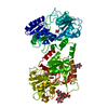
| ||||||||
|---|---|---|---|---|---|---|---|---|---|
| 1 |
| ||||||||
| Unit cell |
|
-Components
-Protein , 1 types, 1 molecules A
| #1: Protein | Mass: 76244.883 Da / Num. of mol.: 1 / Source method: isolated from a natural source / Source: (natural) |
|---|
-Sugars , 3 types, 3 molecules 
| #2: Polysaccharide | beta-D-mannopyranose-(1-4)-2-acetamido-2-deoxy-beta-D-glucopyranose-(1-4)-2-acetamido-2-deoxy-beta- ...beta-D-mannopyranose-(1-4)-2-acetamido-2-deoxy-beta-D-glucopyranose-(1-4)-2-acetamido-2-deoxy-beta-D-glucopyranose Source method: isolated from a genetically manipulated source |
|---|---|
| #3: Polysaccharide | alpha-D-mannopyranose-(1-3)-[alpha-D-mannopyranose-(1-6)]alpha-D-mannopyranose-(1-6)-beta-D- ...alpha-D-mannopyranose-(1-3)-[alpha-D-mannopyranose-(1-6)]alpha-D-mannopyranose-(1-6)-beta-D-mannopyranose-(1-4)-2-acetamido-2-deoxy-beta-D-glucopyranose-(1-4)-2-acetamido-2-deoxy-beta-D-glucopyranose Source method: isolated from a genetically manipulated source |
| #4: Sugar | ChemComp-NAG /  Type: D-saccharide, beta linking / Mass: 221.208 Da / Num. of mol.: 1 Type: D-saccharide, beta linking / Mass: 221.208 Da / Num. of mol.: 1Source method: isolated from a genetically manipulated source Formula: C8H15NO6 |
-Non-polymers , 3 types, 54 molecules 


| #5: Chemical |  Mass: 55.845 Da / Num. of mol.: 2 / Source method: obtained synthetically / Formula: Fe Mass: 55.845 Da / Num. of mol.: 2 / Source method: obtained synthetically / Formula: Fe#6: Chemical |  Mass: 60.009 Da / Num. of mol.: 2 / Source method: obtained synthetically / Formula: CO3 Mass: 60.009 Da / Num. of mol.: 2 / Source method: obtained synthetically / Formula: CO3#7: Water | ChemComp-HOH / | Mass: 18.015 Da / Num. of mol.: 50 / Source method: isolated from a natural source / Formula: H2O |
|---|
-Details
| Has protein modification | Y |
|---|
-Experimental details
-Experiment
| Experiment | Method: X-RAY DIFFRACTION / Number of used crystals: 1 |
|---|
- Sample preparation
| Crystal | Density Matthews: 2.5 Å3/Da / Density % sol: 50 % / Description: USED SEPARATE N AND C LOBES | |||||||||||||||||||||||||
|---|---|---|---|---|---|---|---|---|---|---|---|---|---|---|---|---|---|---|---|---|---|---|---|---|---|---|
| Crystal grow | Temperature: 277 K / Method: dialaysis / pH: 7.7 Details: 25 MM TRIS/HCL, PH 7.7 8% (V/V) MPD 6.5% (V/V) ETHANOL DIALYSED AGAINST A 200 MG/ML SOLUTION OF PROTEIN AT 277 K., dialaysis | |||||||||||||||||||||||||
| Crystal grow | *PLUS Temperature: 4 ℃ / Method: microdialysis | |||||||||||||||||||||||||
| Components of the solutions | *PLUS
|
-Data collection
| Diffraction | Mean temperature: 298 K |
|---|---|
| Diffraction source | Source: ROTATING ANODE / Type: RIGAKU RU300 / Wavelength: 1.5418 |
| Detector | Type: RIGAKU RAXIS IIC / Detector: IMAGE PLATE / Date: May 1, 1994 / Details: 0.3 MM COLLIMATOR |
| Radiation | Monochromator: GRAPHITE(002) / Monochromatic (M) / Laue (L): M / Scattering type: x-ray |
| Radiation wavelength | Wavelength: 1.5418 Å / Relative weight: 1 |
| Reflection | Resolution: 2.8→50 Å / Num. obs: 22017 / % possible obs: 97 % / Observed criterion σ(I): 3 / Redundancy: 3.7 % / Biso Wilson estimate: 62 Å2 / Rmerge(I) obs: 0.048 / Rsym value: 0.048 / Net I/σ(I): 28 |
| Reflection shell | Resolution: 2.8→2.9 Å / Redundancy: 3.7 % / Rmerge(I) obs: 0.31 / Mean I/σ(I) obs: 4 / Rsym value: 0.31 / % possible all: 95.9 |
| Reflection | *PLUS Num. measured all: 81067 |
| Reflection shell | *PLUS % possible obs: 95.9 % |
- Processing
| Software |
| ||||||||||||||||||||||||||||||||||||||||||||||||||
|---|---|---|---|---|---|---|---|---|---|---|---|---|---|---|---|---|---|---|---|---|---|---|---|---|---|---|---|---|---|---|---|---|---|---|---|---|---|---|---|---|---|---|---|---|---|---|---|---|---|---|---|
| Refinement | Method to determine structure: MOLECULAR REPLACEMENT Starting model: HUMAN LACTOFERRIN Resolution: 2.8→30 Å / Isotropic thermal model: BCORREL WITH MODIFICATIONS / Cross valid method: FREE R FACTOR / Stereochemistry target values: PROTGEO WITH MODIFICATIONS
| ||||||||||||||||||||||||||||||||||||||||||||||||||
| Solvent computation | Solvent model: BABINET SCALING / Bsol: 150 Å2 / ksol: 0.8 e/Å3 | ||||||||||||||||||||||||||||||||||||||||||||||||||
| Refinement step | Cycle: LAST / Resolution: 2.8→30 Å
| ||||||||||||||||||||||||||||||||||||||||||||||||||
| Refine LS restraints |
| ||||||||||||||||||||||||||||||||||||||||||||||||||
| Software | *PLUS Name: TNT / Version: 5EA / Classification: refinement | ||||||||||||||||||||||||||||||||||||||||||||||||||
| Refinement | *PLUS Rfactor Rfree: 0.286 | ||||||||||||||||||||||||||||||||||||||||||||||||||
| Solvent computation | *PLUS | ||||||||||||||||||||||||||||||||||||||||||||||||||
| Displacement parameters | *PLUS Biso mean: 68.3 Å2 | ||||||||||||||||||||||||||||||||||||||||||||||||||
| Refine LS restraints | *PLUS
|
