 Movie
Movie Controller


+ Open data

- Basic information
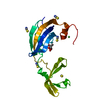
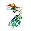
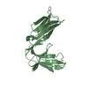
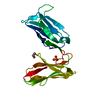
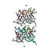
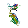
| Entry | Database: PDB / ID: 1nkr | ||||||
|---|---|---|---|---|---|---|---|
| Title | INHIBITORY RECEPTOR (P58-CL42) FOR HUMAN NATURAL KILLER CELLS | ||||||
 Components Components | P58-CL42 KIR | ||||||
 Keywords Keywords | INHIBITORY RECEPTOR / NATURAL KILLER CELLS / IMMUNOLOGICAL RECEPTORS / IMMUNOGLOBULIN FOLD | ||||||
| Function / homology |  Function and homology information Function and homology informationnatural killer cell inhibitory signaling pathway / immune receptor activity / immune response-inhibiting cell surface receptor signaling pathway / Immunoregulatory interactions between a Lymphoid and a non-Lymphoid cell / transmembrane signaling receptor activity / signaling receptor activity / immune response / plasma membrane Similarity search - Function | ||||||
| Biological species |  Homo sapiens (human) Homo sapiens (human) | ||||||
| Method |  X-RAY DIFFRACTION / SYNCHROTRON / MAD / Resolution: 1.7 Å X-RAY DIFFRACTION / SYNCHROTRON / MAD / Resolution: 1.7 Å | ||||||
 Authors Authors | Fan, Q.R. / Mosyak, L. / Winter, C.C. / Wagtmann, N. / Long, E.O. / Wiley, D.C. | ||||||
 Citation Citation | Journal: Nature / Year: 1997 Title: Structure of the inhibitory receptor for human natural killer cells resembles haematopoietic receptors. Authors: Fan, Q.R. / Mosyak, L. / Winter, C.C. / Wagtmann, N. / Long, E.O. / Wiley, D.C. | ||||||
| History |
|
- Structure visualization
| Structure viewer | Molecule: MolmilJmol/JSmol |
|---|
- Downloads & links
-Download
| PDBx/mmCIF format | 1nkr.cif.gz | 55.6 KB | Display | PDBx/mmCIF format |
|---|---|---|---|---|
| PDB format | pdb1nkr.ent.gz | 39.9 KB | Display | PDB format |
| PDBx/mmJSON format | 1nkr.json.gz | Tree view | PDBx/mmJSON format | |
| Others |  Other downloads Other downloads |
-Validation report
| Arichive directory | https://data.pdbj.org/pub/pdb/validation_reports/nk/1nkrftp://data.pdbj.org/pub/pdb/validation_reports/nk/1nkr | HTTPS FTP |
|---|
-Related structure data
| Similar structure data |
|---|


-Links
 PDBj
PDBj


- Assembly
| Deposited unit | 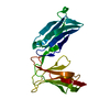
| ||||||||
|---|---|---|---|---|---|---|---|---|---|
| 1 |
| ||||||||
| Unit cell |
|
-Components
| #1: Protein | Mass: 22200.902 Da / Num. of mol.: 1 / Fragment: EXTRACELLULAR DOMAIN Source method: isolated from a genetically manipulated source Source: (gene. exp.) Homo sapiens (human) / Cell: NATURAL KILLER CELLS / Cell line: BL21 / Plasmid: PLM1 / Cellular location (production host): INCLUSION BODY / Production host:  |
|---|---|
| #2: Water | ChemComp-HOH /  Mass: 18.015 Da / Num. of mol.: 211 / Source method: isolated from a natural source / Formula: H2O Mass: 18.015 Da / Num. of mol.: 211 / Source method: isolated from a natural source / Formula: H2O |
| Has protein modification | Y |
-Experimental details
-Experiment
| Experiment | Method: X-RAY DIFFRACTION / Number of used crystals: 1 |
|---|
- Sample preparation
| Crystal | Density Matthews: 1.8 Å3/Da / Density % sol: 40 % | |||||||||||||||
|---|---|---|---|---|---|---|---|---|---|---|---|---|---|---|---|---|
| Crystal grow | pH: 7.7 Details: PROTEIN WAS CRYSTALLIZED FROM 0.55 M (NH4)2HPO4, 50 MM SODIUM CITRATE, PH 5.4, FINAL PH 7.7. CRYSTALS WERE HARVESTED IN 1.5 M (NH4)2HPO4, 50 MM SODIUM CITRATE, PH 5.4, FINAL PH 7.7; THEN ...Details: PROTEIN WAS CRYSTALLIZED FROM 0.55 M (NH4)2HPO4, 50 MM SODIUM CITRATE, PH 5.4, FINAL PH 7.7. CRYSTALS WERE HARVESTED IN 1.5 M (NH4)2HPO4, 50 MM SODIUM CITRATE, PH 5.4, FINAL PH 7.7; THEN SOAKED IN 1.5 M (NH4)2HPO4, 50 MM SODIUM CITRATE, PH 5.4, 25% GLYCEROL, FINAL PH 7.7, AND FLASH-COOLED WITH LIQUID NITROGEN. PH range: 5.4-7.7 | |||||||||||||||
| Crystal | *PLUS | |||||||||||||||
| Crystal grow | *PLUS Method: unknown | |||||||||||||||
| Components of the solutions | *PLUS
|
-Data collection
| Diffraction | Mean temperature: 90 K |
|---|---|
| Diffraction source | Source: SYNCHROTRON / Site: CHESS  / Beamline: F1 / Wavelength: 0.918 / Beamline: F1 / Wavelength: 0.918 |
| Detector | Type: PRINCETON 2K / Detector: CCD / Date: Oct 1, 1996 / Details: MIRRORS |
| Radiation | Monochromator: HORIZONTALLY BENT SI (111), ASYMMETRICALLY CUT CRYSTALS Monochromatic (M) / Laue (L): M / Scattering type: x-ray |
| Radiation wavelength | Wavelength: 0.918 Å / Relative weight: 1 |
| Reflection | Resolution: 1.7→16 Å / Num. obs: 25065 / % possible obs: 99.5 % / Observed criterion σ(I): 0 / Redundancy: 7 % / Rmerge(I) obs: 0.073 / Rsym value: 0.073 / Net I/σ(I): 17.1 |
| Reflection shell | Resolution: 1.7→1.76 Å / Redundancy: 6 % / Rmerge(I) obs: 0.234 / Mean I/σ(I) obs: 4 / Rsym value: 0.234 / % possible all: 97.8 |
| Reflection shell | *PLUS Highest resolution: 1.7 Å / % possible obs: 97.8 % |
- Processing
| Software |
| ||||||||||||||||||||
|---|---|---|---|---|---|---|---|---|---|---|---|---|---|---|---|---|---|---|---|---|---|
| Refinement | Method to determine structure: MAD / Resolution: 1.7→6 Å / Cross valid method: THROUGHOUT / σ(F): 0 Details: THE STRUCTURE WAS INITIALLY REFINED AT 10-2.2 ANGSTROMS USING POSITIONAL REFINEMENT AND SIMULATED ANNEALING PROTOCOLS IN X-PLOR. THE RESOLUTION WAS THEN EXTENDED TO 1.7 ANGSTROMS. REFINEMENT ...Details: THE STRUCTURE WAS INITIALLY REFINED AT 10-2.2 ANGSTROMS USING POSITIONAL REFINEMENT AND SIMULATED ANNEALING PROTOCOLS IN X-PLOR. THE RESOLUTION WAS THEN EXTENDED TO 1.7 ANGSTROMS. REFINEMENT AT THIS STAGE INVOLVED SIMULATED ANNEALING FOLLOWED BY B-FACTOR REFINEMENT, WITH THE EXTENSIVE USE OF SIMULATED ANNEALING OMIT MAPS. THE FINAL MODEL OBTAINED FROM X-PLOR WAS AGAIN REFINED WITH REFMAC. THE FINAL REFINEMENT STATISTICS FROM REFMAC ARE SHOWN HERE.
| ||||||||||||||||||||
| Refinement step | Cycle: LAST / Resolution: 1.7→6 Å
| ||||||||||||||||||||
| Software | *PLUS Name: REFMAC / Classification: refinement | ||||||||||||||||||||
| Refine LS restraints | *PLUS
|
