 Movie
Movie Controller

[English] 日本語
 Yorodumi
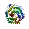
- PDB-1nc5: Structure of Protein of Unknown Function of YteR from Bacillus Su... -
+ Open data

- Basic information
| Entry | Database: PDB / ID: 1nc5 | ||||||
|---|---|---|---|---|---|---|---|
| Title | Structure of Protein of Unknown Function of YteR from Bacillus Subtilis | ||||||
 Components Components | hypothetical protein yTER | ||||||
 Keywords Keywords | STRUCTURAL GENOMICS / UNKNOWN FUNCTION / HELIX BARREL / PSI / Protein Structure Initiative / Midwest Center for Structural Genomics / MCSG | ||||||
| Function / homology |  Function and homology information Function and homology informationunsaturated rhamnogalacturonyl hydrolase / unsaturated rhamnogalacturonyl hydrolase activity / carbohydrate metabolic process / cytoplasm Similarity search - Function | ||||||
| Biological species |  | ||||||
| Method |  X-RAY DIFFRACTION / SYNCHROTRON / MAD / Resolution: 1.6 Å X-RAY DIFFRACTION / SYNCHROTRON / MAD / Resolution: 1.6 Å | ||||||
 Authors Authors | Zhang, R. / Lozondra, L. / Korolev, S. / Collart, F. / Joachimiak, A. / Midwest Center for Structural Genomics (MCSG) | ||||||
 Citation Citation | Journal: Proteins / Year: 2005 Title: 1.6 A crystal structure of YteR protein from Bacillus subtilis, a predicted lyase. Authors: Zhang, R. / Minh, T. / Lezondra, L. / Korolev, S. / Moy, S.F. / Collart, F. / Joachimiak, A. | ||||||
| History |
|
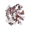
- Structure visualization
| Structure viewer | Molecule: MolmilJmol/JSmol |
|---|
- Downloads & links
-Download
| PDBx/mmCIF format | 1nc5.cif.gz | 95.8 KB | Display | PDBx/mmCIF format |
|---|---|---|---|---|
| PDB format | pdb1nc5.ent.gz | 72.5 KB | Display | PDB format |
| PDBx/mmJSON format | 1nc5.json.gz | Tree view | PDBx/mmJSON format | |
| Others |  Other downloads Other downloads |
-Validation report
| Summary document | 1nc5_validation.pdf.gz | 366.8 KB | Display | wwPDB validaton report |
|---|---|---|---|---|
| Full document | 1nc5_full_validation.pdf.gz | 369.7 KB | Display | |
| Data in XML | 1nc5_validation.xml.gz | 8.4 KB | Display | |
| Data in CIF | 1nc5_validation.cif.gz | 15.2 KB | Display | |
| Arichive directory | https://data.pdbj.org/pub/pdb/validation_reports/nc/1nc5ftp://data.pdbj.org/pub/pdb/validation_reports/nc/1nc5 | HTTPS FTP |
-Related structure data
| Similar structure data | |
|---|---|
| Other databases |


-Links
 PDBj
PDBj
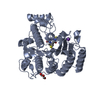
- Assembly
| Deposited unit | 
| ||||||||
|---|---|---|---|---|---|---|---|---|---|
| 1 |
| ||||||||
| Unit cell |
| ||||||||
| Details | This protein (APC1644) existed as monomer |
-Components
| #1: Protein | Mass: 43021.707 Da / Num. of mol.: 1 Source method: isolated from a genetically manipulated source Source: (gene. exp.) |
|---|---|
| #2: Water | ChemComp-HOH /  Mass: 18.015 Da / Num. of mol.: 499 / Source method: isolated from a natural source / Formula: H2O Mass: 18.015 Da / Num. of mol.: 499 / Source method: isolated from a natural source / Formula: H2O |
-Experimental details
-Experiment
| Experiment | Method: X-RAY DIFFRACTION / Number of used crystals: 1 |
|---|
- Sample preparation
| Crystal | Density Matthews: 2.54 Å3/Da / Density % sol: 51.56 % | ||||||||||||||||||||||||
|---|---|---|---|---|---|---|---|---|---|---|---|---|---|---|---|---|---|---|---|---|---|---|---|---|---|
| Crystal grow | Temperature: 298 K / Method: vapor diffusion, hanging drop / pH: 7.5 Details: 1.3 M Nacitrate, 0.1M Tris , pH 7.5, VAPOR DIFFUSION, HANGING DROP, temperature 298K | ||||||||||||||||||||||||
| Crystal grow | *PLUS Temperature: 295 K / Method: vapor diffusion, hanging drop | ||||||||||||||||||||||||
| Components of the solutions | *PLUS
|
-Data collection
| Diffraction | Mean temperature: 100 K | ||||||||||||
|---|---|---|---|---|---|---|---|---|---|---|---|---|---|
| Diffraction source | Source: SYNCHROTRON / Site: APS  / Beamline: 19-ID / Wavelength: 0.9795,0.9798,0.94656 / Beamline: 19-ID / Wavelength: 0.9795,0.9798,0.94656 | ||||||||||||
| Detector | Type: SBC-2 / Detector: CCD / Date: Nov 12, 2002 / Details: mirrors | ||||||||||||
| Radiation | Monochromator: Si 111 CHANNEL / Protocol: MAD / Monochromatic (M) / Laue (L): M / Scattering type: x-ray | ||||||||||||
| Radiation wavelength |
| ||||||||||||
| Reflection | Resolution: 1.6→50 Å / Num. obs: 59605 / % possible obs: 99.3 % / Observed criterion σ(F): 4 / Observed criterion σ(I): 4 / Redundancy: 20.36 % / Biso Wilson estimate: 18.4 Å2 / Rmerge(I) obs: 0.102 / Net I/σ(I): 34.88 | ||||||||||||
| Reflection shell | Resolution: 1.6→1.66 Å / Redundancy: 5.6 % / Rmerge(I) obs: 0.561 / Mean I/σ(I) obs: 2.47 / Num. unique all: 5741 / % possible all: 98.3 | ||||||||||||
| Reflection | *PLUS Highest resolution: 1.6 Å / Num. obs: 59188 | ||||||||||||
| Reflection shell | *PLUS % possible obs: 98.3 % / Rmerge(I) obs: 0.56 |
- Processing
| Software |
| ||||||||||||||||||||||||||||||||||||
|---|---|---|---|---|---|---|---|---|---|---|---|---|---|---|---|---|---|---|---|---|---|---|---|---|---|---|---|---|---|---|---|---|---|---|---|---|---|
| Refinement | Method to determine structure: MAD Starting model: none Resolution: 1.6→44.49 Å / Rfactor Rfree error: 0.003 / Isotropic thermal model: RESTRAINED / Cross valid method: THROUGHOUT / σ(F): 0 / Stereochemistry target values: Engh & Huber Details: In the cns refinement (hlml target), the Friedel's pair was treated as two seperated reflections.
| ||||||||||||||||||||||||||||||||||||
| Solvent computation | Solvent model: FLAT MODEL / Bsol: 60.3677 Å2 / ksol: 0.389443 e/Å3 | ||||||||||||||||||||||||||||||||||||
| Displacement parameters | Biso mean: 20 Å2
| ||||||||||||||||||||||||||||||||||||
| Refine analyze | Luzzati coordinate error free: 0.2 Å / Luzzati sigma a free: 0.15 Å | ||||||||||||||||||||||||||||||||||||
| Refinement step | Cycle: LAST / Resolution: 1.6→44.49 Å
| ||||||||||||||||||||||||||||||||||||
| Refine LS restraints |
| ||||||||||||||||||||||||||||||||||||
| LS refinement shell | Resolution: 1.6→1.7 Å / Rfactor Rfree error: 0.01 / Total num. of bins used: 6
| ||||||||||||||||||||||||||||||||||||
| Xplor file |
| ||||||||||||||||||||||||||||||||||||
| Refine LS restraints | *PLUS
|
