 Movie
Movie Controller

[English] 日本語
 Yorodumi
- PDB-2zun: Functional Analysis of Hyperthermophilic Endocellulase from the A... -
+ Open data

- Basic information
| Entry | Database: PDB / ID: 2zun | |||||||||
|---|---|---|---|---|---|---|---|---|---|---|
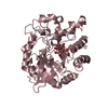
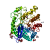
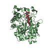
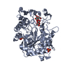
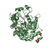
| Title | Functional Analysis of Hyperthermophilic Endocellulase from the Archaeon Pyrococcus horikoshii | |||||||||
 Components Components | 458aa long hypothetical endo-1,4-beta-glucanase | |||||||||
 Keywords Keywords | HYDROLASE / TIM BARREL | |||||||||
| Function / homology |  Function and homology information Function and homology informationhydrolase activity, hydrolyzing O-glycosyl compounds / cellulose catabolic process / metal ion binding Similarity search - Function | |||||||||
| Biological species |   Pyrococcus horikoshii (archaea) Pyrococcus horikoshii (archaea) | |||||||||
| Method |  X-RAY DIFFRACTION / SYNCHROTRON / MOLECULAR REPLACEMENT / Resolution: 2 Å X-RAY DIFFRACTION / SYNCHROTRON / MOLECULAR REPLACEMENT / Resolution: 2 Å | |||||||||
 Authors Authors | Kim, H.-W. / Ishikawa, K. | |||||||||
 Citation Citation | Journal: To be Published Title: Functional Analysis of Hyperthermophilic Endocellulase from the Archaeon Pyrococcus horikoshii Authors: Kim, H.-W. / Ishikawa, K. | |||||||||
| History |
|
- Structure visualization
| Structure viewer | Molecule: MolmilJmol/JSmol |
|---|
- Downloads & links
-Download
| PDBx/mmCIF format | 2zun.cif.gz | 251.3 KB | Display | PDBx/mmCIF format |
|---|---|---|---|---|
| PDB format | pdb2zun.ent.gz | 202.7 KB | Display | PDB format |
| PDBx/mmJSON format | 2zun.json.gz | Tree view | PDBx/mmJSON format | |
| Others |  Other downloads Other downloads |
-Validation report
| Arichive directory | https://data.pdbj.org/pub/pdb/validation_reports/zu/2zunftp://data.pdbj.org/pub/pdb/validation_reports/zu/2zun | HTTPS FTP |
|---|
-Related structure data
| Related structure data |  2zumS S: Starting model for refinement |
|---|---|
| Similar structure data |


-Links
 PDBj
PDBj- Assembly
| Deposited unit | 
| ||||||||
|---|---|---|---|---|---|---|---|---|---|
| 1 | 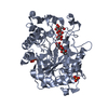
| ||||||||
| 2 | 
| ||||||||
| 3 | 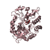
| ||||||||
| Unit cell |
|
-Components
| #1: Protein | Mass: 51954.863 Da / Num. of mol.: 3 Source method: isolated from a genetically manipulated source Source: (gene. exp.) Pyrococcus horikoshii (archaea) / Strain: OT3 / Gene: EGPh / Plasmid: pET11a / Production host:  #2: Polysaccharide | beta-D-glucopyranose-(1-4)-beta-D-glucopyranose / beta-cellobiose   Source method: isolated from a genetically manipulated source Details: oligosaccharide / References: beta-cellobiose #3: Chemical |   Mass: 94.971 Da / Num. of mol.: 3 / Source method: obtained synthetically / Formula: PO4 Mass: 94.971 Da / Num. of mol.: 3 / Source method: obtained synthetically / Formula: PO4#4: Water | ChemComp-HOH / |  Mass: 18.015 Da / Num. of mol.: 486 / Source method: isolated from a natural source / Formula: H2O Mass: 18.015 Da / Num. of mol.: 486 / Source method: isolated from a natural source / Formula: H2OHas protein modification | Y | Sequence details | THERE IS CONFLICT BETWEEN THE REPORTED SEQUENCE AND THE DATABASE REFERENCE SEQUENCE. IN THIS ENTRY, ...THERE IS CONFLICT BETWEEN THE REPORTED SEQUENCE AND THE DATABASE REFERENCE SEQUENCE. IN THIS ENTRY, 289TH RESIDUE IS OBVIOUSLY LYS BASED ON THE ELECTRON DENSITY. | |
|---|
-Experimental details
-Experiment
| Experiment | Method: X-RAY DIFFRACTION / Number of used crystals: 1 |
|---|
- Sample preparation
| Crystal | Density Matthews: 1.98 Å3/Da / Density % sol: 38.01 % |
|---|---|
| Crystal grow | Temperature: 295 K / Method: vapor diffusion, hanging drop / pH: 6.5 Details: 1.5M ammonium phosphate, 0.1M MES buffer, pH 6.5, VAPOR DIFFUSION, HANGING DROP, temperature 295K |
-Data collection
| Diffraction | Mean temperature: 100 K |
|---|---|
| Diffraction source | Source: SYNCHROTRON / Site: SPring-8  / Beamline: BL44XU / Wavelength: 0.9 Å / Beamline: BL44XU / Wavelength: 0.9 Å |
| Detector | Type: Bruker DIP-6040 / Detector: CCD |
| Radiation | Protocol: SINGLE WAVELENGTH / Monochromatic (M) / Laue (L): M / Scattering type: x-ray |
| Radiation wavelength | Wavelength: 0.9 Å / Relative weight: 1 |
| Reflection | Resolution: 1.9→50 Å / Num. all: 97064 / Num. obs: 94740 / % possible obs: 97.6 % / Redundancy: 3.5 % |
- Processing
| Software |
| |||||||||||||||||||||
|---|---|---|---|---|---|---|---|---|---|---|---|---|---|---|---|---|---|---|---|---|---|---|
| Refinement | Method to determine structure: MOLECULAR REPLACEMENT Starting model: PDB ENTRY 2ZUM Resolution: 2→48.37 Å / Cor.coef. Fo:Fc: 0.957 / Cor.coef. Fo:Fc free: 0.921 / Occupancy max: 1 / Occupancy min: 0.5 / SU B: 5.385 / SU ML: 0.148 / Cross valid method: THROUGHOUT / σ(F): 0 / ESU R: 0.208 / ESU R Free: 0.196 / Stereochemistry target values: MAXIMUM LIKELIHOOD / Details: HYDROGENS HAVE BEEN ADDED IN THE RIDING POSITIONS
| |||||||||||||||||||||
| Solvent computation | Ion probe radii: 0.8 Å / Shrinkage radii: 0.8 Å / VDW probe radii: 1.2 Å / Solvent model: MASK / Bsol: 73.825 Å2 | |||||||||||||||||||||
| Displacement parameters | Biso max: 84.75 Å2 / Biso mean: 37.085 Å2 / Biso min: 9.12 Å2
| |||||||||||||||||||||
| Refinement step | Cycle: LAST / Resolution: 2→48.37 Å
| |||||||||||||||||||||
| Refine LS restraints |
| |||||||||||||||||||||
| LS refinement shell | Resolution: 2→2.052 Å / Total num. of bins used: 20
| |||||||||||||||||||||
| Xplor file |
|
