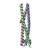
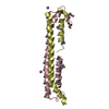
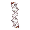
- PDB-1naf: Crystal structure of the human GGA1 GAT domain -
+
データを開く
IDまたはキーワード:
読み込み中...
-
基本情報
登録情報
データベース: PDB / ID: 1naf
タイトル
Crystal structure of the human GGA1 GAT domain
要素
ADP-ribosylation factor binding protein GGA1
キーワード
SIGNALING PROTEIN / MEMBRANE PROTEIN / clathrin-adaptor / GGA / GAT domain / helical paper-clip / three-helix bundle
機能・相同性
機能・相同性情報
protein localization to ciliary membrane / Golgi to plasma membrane transport / Golgi to plasma membrane protein transport / TBC/RABGAPs / protein localization to cell surface / retrograde transport, endosome to Golgi / phosphatidylinositol binding / protein catabolic process / ubiquitin binding / trans-Golgi network ...protein localization to ciliary membrane / Golgi to plasma membrane transport / Golgi to plasma membrane protein transport / TBC/RABGAPs / protein localization to cell surface / retrograde transport, endosome to Golgi / phosphatidylinositol binding / protein catabolic process / ubiquitin binding / trans-Golgi network / intracellular protein transport / small GTPase binding / positive regulation of protein catabolic process / intracellular protein localization / early endosome membrane / early endosome / endosome membrane / Amyloid fiber formation / Golgi apparatus / protein-containing complex / nucleoplasm / membrane / cytosol 類似検索 - 分子機能
Methane Monooxygenase Hydroxylase; Chain G, domain 1 - #160 / ADP-ribosylation factor-binding protein GGA3 / N-terminal extension of GAT domain / N-terminal extension of GAT domain / GAT domain / GAT domain superfamily / GAT domain / GAT domain profile. / VHS domain / VHS domain ...Methane Monooxygenase Hydroxylase; Chain G, domain 1 - #160 / ADP-ribosylation factor-binding protein GGA3 / N-terminal extension of GAT domain / N-terminal extension of GAT domain / GAT domain / GAT domain superfamily / GAT domain / GAT domain profile. / VHS domain / VHS domain / VHS domain profile. / Domain present in VPS-27, Hrs and STAM / Gamma-adaptin ear (GAE) domain / Gamma-adaptin ear (GAE) domain profile. / Clathrin adaptor, alpha/beta/gamma-adaptin, appendage, Ig-like subdomain / Adaptin C-terminal domain / Adaptin C-terminal domain / Clathrin adaptor, appendage, Ig-like subdomain superfamily / ENTH/VHS / Single alpha-helices involved in coiled-coils or other helix-helix interfaces - #170 / Single alpha-helices involved in coiled-coils or other helix-helix interfaces / Methane Monooxygenase Hydroxylase; Chain G, domain 1 / Up-down Bundle / Mainly Alpha 類似検索 - ドメイン・相同性
ADP-ribosylation factor-binding protein GGA1 類似検索 - 構成要素
 ムービー
ムービー コントローラー


 データを開く
 基本情報
基本情報 要素
要素 キーワード
キーワード 機能・相同性情報
 Homo sapiens (ヒト)
 X線回折 /
  データ登録者
 引用
引用 構造の表示
構造の表示 ダウンロードとリンク
 その他のダウンロード


 PDBj
PDBj


 集合体
集合体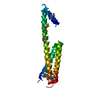

 試料調製
試料調製 / ビームライン: ID14-4 / 波長: 0.9796, 0.9798, 0.9393
 解析
解析