 Movie
Movie Controller

[English] 日本語
 Yorodumi
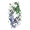
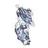
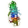
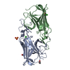
- PDB-1na8: Crystal structure of ADP-ribosylation factor binding protein GGA1 -
+ Open data

- Basic information
| Entry | Database: PDB / ID: 1na8 | ||||||
|---|---|---|---|---|---|---|---|
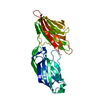
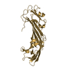
| Title | Crystal structure of ADP-ribosylation factor binding protein GGA1 | ||||||
 Components Components | ADP-ribosylation factor binding protein GGA1 | ||||||
 Keywords Keywords | SIGNALING PROTEIN / MEMBRANE PROTEIN / clathrin-adaptor / GGA / appendage / beta-sandwich | ||||||
| Function / homology |  Function and homology information Function and homology informationprotein localization to ciliary membrane / Golgi to plasma membrane transport / Golgi to plasma membrane protein transport / TBC/RABGAPs / retrograde transport, endosome to Golgi / protein localization to cell surface / phosphatidylinositol binding / ubiquitin binding / trans-Golgi network / intracellular protein transport ...protein localization to ciliary membrane / Golgi to plasma membrane transport / Golgi to plasma membrane protein transport / TBC/RABGAPs / retrograde transport, endosome to Golgi / protein localization to cell surface / phosphatidylinositol binding / ubiquitin binding / trans-Golgi network / intracellular protein transport / small GTPase binding / intracellular protein localization / early endosome membrane / early endosome / endosome membrane / Amyloid fiber formation / Golgi apparatus / protein-containing complex / membrane / cytosol Similarity search - Function | ||||||
| Biological species |  Homo sapiens (human) Homo sapiens (human) | ||||||
| Method |  X-RAY DIFFRACTION / MOLECULAR REPLACEMENT / Resolution: 2.3 Å X-RAY DIFFRACTION / MOLECULAR REPLACEMENT / Resolution: 2.3 Å | ||||||
 Authors Authors | Lui, W.W. / Collins, B.M. / Hirst, J. / Motley, A. / Millar, C. / Schu, P. / Owen, D.J. / Robinson, M.S. | ||||||
 Citation Citation | Journal: Mol.Cell.Biol. / Year: 2003 Title: Binding partners for the COOH-terminal appendage domains of the GGAs and gamma-adaptin Authors: Lui, W.W. / Collins, B.M. / Hirst, J. / Motley, A. / Millar, C. / Schu, P. / Owen, D.J. / Robinson, M.S. | ||||||
| History |
|
- Structure visualization
| Structure viewer | Molecule: MolmilJmol/JSmol |
|---|
- Downloads & links
-Download
| PDBx/mmCIF format | 1na8.cif.gz | 73 KB | Display | PDBx/mmCIF format |
|---|---|---|---|---|
| PDB format | pdb1na8.ent.gz | 55.2 KB | Display | PDB format |
| PDBx/mmJSON format | 1na8.json.gz | Tree view | PDBx/mmJSON format | |
| Others |  Other downloads Other downloads |
-Validation report
| Arichive directory | https://data.pdbj.org/pub/pdb/validation_reports/na/1na8ftp://data.pdbj.org/pub/pdb/validation_reports/na/1na8 | HTTPS FTP |
|---|
-Related structure data
| Related structure data |  1gyuS S: Starting model for refinement |
|---|---|
| Similar structure data |


-Links
 PDBj
PDBj


- Assembly
| Deposited unit | 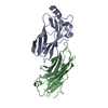
| ||||||||||
|---|---|---|---|---|---|---|---|---|---|---|---|
| 1 |
| ||||||||||
| Unit cell |
| ||||||||||
| Details | The assymetric unit contains a dimer of two identical chains but this dimer is not observed in solution |
-Components
| #1: Protein | Mass: 17353.209 Da / Num. of mol.: 2 / Fragment: appendage domain, Residues 494-639 of SWS Q9UJY5 Source method: isolated from a genetically manipulated source Source: (gene. exp.) Homo sapiens (human) / Plasmid: pET / Production host:  #2: Water | ChemComp-HOH / |  Mass: 18.015 Da / Num. of mol.: 172 / Source method: isolated from a natural source / Formula: H2O Mass: 18.015 Da / Num. of mol.: 172 / Source method: isolated from a natural source / Formula: H2O |
|---|
-Experimental details
-Experiment
| Experiment | Method: X-RAY DIFFRACTION / Number of used crystals: 1 |
|---|
- Sample preparation
| Crystal | Density Matthews: 2.52 Å3/Da / Density % sol: 50.8 % | ||||||||||||||||||||||||
|---|---|---|---|---|---|---|---|---|---|---|---|---|---|---|---|---|---|---|---|---|---|---|---|---|---|
| Crystal grow | Temperature: 289 K / Method: vapor diffusion, sitting drop / pH: 5.6 Details: sodium citrate, ammonium sulphate, pH 5.6, VAPOR DIFFUSION, SITTING DROP, temperature 289K | ||||||||||||||||||||||||
| Crystal grow | *PLUS Method: vapor diffusion, sitting drop | ||||||||||||||||||||||||
| Components of the solutions | *PLUS
|
-Data collection
| Diffraction | Mean temperature: 100 K |
|---|---|
| Diffraction source | Source: ROTATING ANODE / Type: RIGAKU / Wavelength: 1.5418 Å |
| Detector | Type: MARRESEARCH / Detector: IMAGE PLATE / Date: 2001 |
| Radiation | Protocol: SINGLE WAVELENGTH / Monochromatic (M) / Laue (L): M / Scattering type: x-ray |
| Radiation wavelength | Wavelength: 1.5418 Å / Relative weight: 1 |
| Reflection | Resolution: 2.3→57 Å / Num. obs: 16193 / % possible obs: 99 % / Observed criterion σ(F): 5.9 / Observed criterion σ(I): -3 / Redundancy: 5.6 % / Biso Wilson estimate: 45 Å2 / Rmerge(I) obs: 0.053 / Net I/σ(I): 23.7 |
| Reflection shell | Resolution: 2.3→2.36 Å / Redundancy: 5.7 % / Rmerge(I) obs: 0.306 / Mean I/σ(I) obs: 5.9 / % possible all: 99 |
| Reflection | *PLUS Highest resolution: 2.3 Å / Lowest resolution: 57 Å / % possible obs: 99 % |
| Reflection shell | *PLUS % possible obs: 99 % |
- Processing
| Software |
| ||||||||||||||||||||||||||||||||||||||||||||||||||||||||||||||||||||||||||||||||||||||||||||||||||||||||||||||||||||||||||||||||||
|---|---|---|---|---|---|---|---|---|---|---|---|---|---|---|---|---|---|---|---|---|---|---|---|---|---|---|---|---|---|---|---|---|---|---|---|---|---|---|---|---|---|---|---|---|---|---|---|---|---|---|---|---|---|---|---|---|---|---|---|---|---|---|---|---|---|---|---|---|---|---|---|---|---|---|---|---|---|---|---|---|---|---|---|---|---|---|---|---|---|---|---|---|---|---|---|---|---|---|---|---|---|---|---|---|---|---|---|---|---|---|---|---|---|---|---|---|---|---|---|---|---|---|---|---|---|---|---|---|---|---|---|
| Refinement | Method to determine structure: MOLECULAR REPLACEMENT Starting model: PDB ID 1GYU Resolution: 2.3→15 Å / Cor.coef. Fo:Fc: 0.939 / Cor.coef. Fo:Fc free: 0.906 / TLS residual ADP flag: LIKELY RESIDUAL / Isotropic thermal model: TLS refinement used / Cross valid method: THROUGHOUT / σ(F): 0 / Stereochemistry target values: MAXIMUM LIKELIHOOD / Details: HYDROGENS HAVE BEEN ADDED IN THE RIDING POSITIONS
| ||||||||||||||||||||||||||||||||||||||||||||||||||||||||||||||||||||||||||||||||||||||||||||||||||||||||||||||||||||||||||||||||||
| Solvent computation | Shrinkage radii: 0.8 Å / Solvent model: BABINET MODEL WITH MASK | ||||||||||||||||||||||||||||||||||||||||||||||||||||||||||||||||||||||||||||||||||||||||||||||||||||||||||||||||||||||||||||||||||
| Displacement parameters | Biso mean: 23.119 Å2
| ||||||||||||||||||||||||||||||||||||||||||||||||||||||||||||||||||||||||||||||||||||||||||||||||||||||||||||||||||||||||||||||||||
| Refinement step | Cycle: LAST / Resolution: 2.3→15 Å
| ||||||||||||||||||||||||||||||||||||||||||||||||||||||||||||||||||||||||||||||||||||||||||||||||||||||||||||||||||||||||||||||||||
| Refine LS restraints |
| ||||||||||||||||||||||||||||||||||||||||||||||||||||||||||||||||||||||||||||||||||||||||||||||||||||||||||||||||||||||||||||||||||
| LS refinement shell | Resolution: 2.3→2.358 Å / Total num. of bins used: 20 /
| ||||||||||||||||||||||||||||||||||||||||||||||||||||||||||||||||||||||||||||||||||||||||||||||||||||||||||||||||||||||||||||||||||
| Refinement TLS params. | Method: refined / Refine-ID: X-RAY DIFFRACTION
| ||||||||||||||||||||||||||||||||||||||||||||||||||||||||||||||||||||||||||||||||||||||||||||||||||||||||||||||||||||||||||||||||||
| Refinement TLS group |
| ||||||||||||||||||||||||||||||||||||||||||||||||||||||||||||||||||||||||||||||||||||||||||||||||||||||||||||||||||||||||||||||||||
| Refinement | *PLUS Highest resolution: 2.3 Å / Lowest resolution: 15 Å / Rfactor Rfree: 0.279 / Rfactor Rwork: 0.219 | ||||||||||||||||||||||||||||||||||||||||||||||||||||||||||||||||||||||||||||||||||||||||||||||||||||||||||||||||||||||||||||||||||
| Solvent computation | *PLUS | ||||||||||||||||||||||||||||||||||||||||||||||||||||||||||||||||||||||||||||||||||||||||||||||||||||||||||||||||||||||||||||||||||
| Displacement parameters | *PLUS | ||||||||||||||||||||||||||||||||||||||||||||||||||||||||||||||||||||||||||||||||||||||||||||||||||||||||||||||||||||||||||||||||||
| Refine LS restraints | *PLUS
| ||||||||||||||||||||||||||||||||||||||||||||||||||||||||||||||||||||||||||||||||||||||||||||||||||||||||||||||||||||||||||||||||||
| LS refinement shell | *PLUS Highest resolution: 2.3 Å / Lowest resolution: 2.36 Å |
