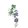
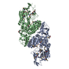
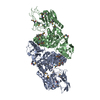
 Movie
Movie Controller


+ Open data

- Basic information
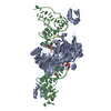
| Entry | Database: PDB / ID: 1n8y | |||||||||
|---|---|---|---|---|---|---|---|---|---|---|
| Title | Crystal structure of the extracellular region of rat HER2 | |||||||||
 Components Components | protooncoprotein | |||||||||
 Keywords Keywords | TRANSFERASE / tyrosin kinase receptor / cell surface receptor | |||||||||
| Function / homology |  Function and homology information Function and homology informationSignaling by ERBB2 / GRB7 events in ERBB2 signaling / ERBB2 Activates PTK6 Signaling / Drug-mediated inhibition of ERBB2 signaling / GRB2 events in ERBB2 signaling / ERBB2 Regulates Cell Motility / PI3K events in ERBB2 signaling / Downregulation of ERBB2:ERBB3 signaling / Downregulation of ERBB2 signaling / Sema4D induced cell migration and growth-cone collapse ...Signaling by ERBB2 / GRB7 events in ERBB2 signaling / ERBB2 Activates PTK6 Signaling / Drug-mediated inhibition of ERBB2 signaling / GRB2 events in ERBB2 signaling / ERBB2 Regulates Cell Motility / PI3K events in ERBB2 signaling / Downregulation of ERBB2:ERBB3 signaling / Downregulation of ERBB2 signaling / Sema4D induced cell migration and growth-cone collapse / SHC1 events in ERBB2 signaling / positive regulation of membrane permeability / PIP3 activates AKT signaling / tongue development / negative regulation of immature T cell proliferation in thymus / PI5P, PP2A and IER3 Regulate PI3K/AKT Signaling / mammary gland involution / ERBB3:ERBB2 complex / ERBB2-ERBB4 signaling pathway / lateral loop / sympathetic nervous system development / RNA polymerase I core binding / glial cell differentiation / semaphorin receptor complex / peripheral nervous system development / ErbB-3 class receptor binding / positive regulation of Ras protein signal transduction / motor neuron axon guidance / positive regulation of neural precursor cell proliferation / regulation of microtubule-based process / RAF/MAP kinase cascade / response to vitamin D / axon regeneration / ERBB signaling pathway / ERBB2-EGFR signaling pathway / response to dexamethasone / response to muscle activity / ERBB2-ERBB3 signaling pathway / neurotransmitter receptor localization to postsynaptic specialization membrane / positive regulation of Rho protein signal transduction / positive regulation of MAP kinase activity / oligodendrocyte differentiation / positive regulation of transcription by RNA polymerase I / microvillus / semaphorin-plexin signaling pathway / regulation of cell differentiation / neuromuscular junction development / multicellular organismal response to stress / response to axon injury / positive regulation of protein targeting to membrane / regulation of ERK1 and ERK2 cascade / Schwann cell development / skeletal muscle tissue development / estrous cycle / coreceptor activity / cell projection / response to progesterone / myelination / peptidyl-tyrosine phosphorylation / transmembrane receptor protein tyrosine kinase activity / positive regulation of epithelial cell proliferation / positive regulation of cell adhesion / cell surface receptor protein tyrosine kinase signaling pathway / cellular response to epidermal growth factor stimulus / basal plasma membrane / liver development / positive regulation of translation / phosphatidylinositol 3-kinase/protein kinase B signal transduction / neuromuscular junction / wound healing / female pregnancy / cellular response to mechanical stimulus / receptor protein-tyrosine kinase / Hsp90 protein binding / cellular response to growth factor stimulus / receptor tyrosine kinase binding / positive regulation of JNK cascade / epidermal growth factor receptor signaling pathway / ruffle membrane / neuron differentiation / transmembrane signaling receptor activity / protein autophosphorylation / myelin sheath / regulation of cell population proliferation / nervous system development / heart development / positive regulation of cell growth / protein tyrosine kinase activity / presynaptic membrane / cytoplasmic vesicle / basolateral plasma membrane / positive regulation of MAPK cascade / early endosome / positive regulation of phosphatidylinositol 3-kinase/protein kinase B signal transduction / cell population proliferation / postsynaptic membrane / cell surface receptor signaling pathway / signaling receptor complex / apical plasma membrane / endosome membrane Similarity search - Function | |||||||||
| Biological species |  | |||||||||
| Method |  X-RAY DIFFRACTION / SYNCHROTRON / MOLECULAR REPLACEMENT / Resolution: 2.4 Å X-RAY DIFFRACTION / SYNCHROTRON / MOLECULAR REPLACEMENT / Resolution: 2.4 Å | |||||||||
 Authors Authors | Cho, H.-S. / Mason, K. / Ramyar, K.X. / Stanley, A.M. / Gabelli, S.B. / Denney Jr., D.W. / Leahy, D.J. | |||||||||
 Citation Citation | Journal: Nature / Year: 2003 Title: Structure of the Extracellular Region of HER2 Alone and in complex with the Herceptin Fab Authors: Cho, H.-S. / Mason, K. / Ramyar, K.X. / Stanley, A.M. / Gabelli, S.B. / Denney Jr., D.W. / Leahy, D.J. | |||||||||
| History |
|
- Structure visualization
| Structure viewer | Molecule: MolmilJmol/JSmol |
|---|
- Downloads & links
-Download
| PDBx/mmCIF format | 1n8y.cif.gz | 130.2 KB | Display | PDBx/mmCIF format |
|---|---|---|---|---|
| PDB format | pdb1n8y.ent.gz | 99.8 KB | Display | PDB format |
| PDBx/mmJSON format | 1n8y.json.gz | Tree view | PDBx/mmJSON format | |
| Others |  Other downloads Other downloads |
-Validation report
| Arichive directory | https://data.pdbj.org/pub/pdb/validation_reports/n8/1n8yftp://data.pdbj.org/pub/pdb/validation_reports/n8/1n8y | HTTPS FTP |
|---|
-Related structure data
| Related structure data | 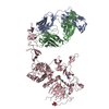 1n8zC 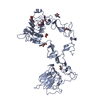 1m6bS S: Starting model for refinement C: citing same article ( |
|---|---|
| Similar structure data |


-Links
 PDBj
PDBj


- Assembly
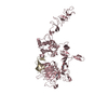
| Deposited unit | 
| ||||||||
|---|---|---|---|---|---|---|---|---|---|
| 1 |
| ||||||||
| Unit cell |
|
-Components
| #1: Protein | Mass: 67361.422 Da / Num. of mol.: 1 / Fragment: extracellular region (residues 26-633) Source method: isolated from a genetically manipulated source Source: (gene. exp.)  Cricetulus griseus (Chinese hamster) / Strain (production host): CHO(Lec1) / References: GenBank: 22651765, UniProt: P06494*PLUS Cricetulus griseus (Chinese hamster) / Strain (production host): CHO(Lec1) / References: GenBank: 22651765, UniProt: P06494*PLUS | ||||||
|---|---|---|---|---|---|---|---|
| #2: Polysaccharide | Source method: isolated from a genetically manipulated source #3: Sugar | ChemComp-NAG / |   Type: D-saccharide, beta linking / Mass: 221.208 Da / Num. of mol.: 1 Type: D-saccharide, beta linking / Mass: 221.208 Da / Num. of mol.: 1Source method: isolated from a genetically manipulated source Formula: C8H15NO6 #4: Water | ChemComp-HOH / |  Mass: 18.015 Da / Num. of mol.: 84 / Source method: isolated from a natural source / Formula: H2O Mass: 18.015 Da / Num. of mol.: 84 / Source method: isolated from a natural source / Formula: H2OHas protein modification | Y | |
-Experimental details
-Experiment
| Experiment | Method: X-RAY DIFFRACTION / Number of used crystals: 1 |
|---|
- Sample preparation
| Crystal | Density Matthews: 3.18 Å3/Da / Density % sol: 61.05 % | ||||||||||||||||||||||||||||||||||||
|---|---|---|---|---|---|---|---|---|---|---|---|---|---|---|---|---|---|---|---|---|---|---|---|---|---|---|---|---|---|---|---|---|---|---|---|---|---|
| Crystal grow | Temperature: 298 K / Method: vapor diffusion, hanging drop / pH: 5.4 Details: 15-30% PEG4000, 50mM Na citrate pH5.4, 10mM EDTA, VAPOR DIFFUSION, HANGING DROP, temperature 298K | ||||||||||||||||||||||||||||||||||||
| Crystal grow | *PLUS pH: 7.5 | ||||||||||||||||||||||||||||||||||||
| Components of the solutions | *PLUS
|
-Data collection
| Diffraction | Mean temperature: 110 K |
|---|---|
| Diffraction source | Source: SYNCHROTRON / Site: NSLS  / Beamline: X4A / Beamline: X4A |
| Detector | Type: ADSC QUANTUM 4 / Detector: CCD / Date: Feb 20, 2001 |
| Radiation | Protocol: SINGLE WAVELENGTH / Monochromatic (M) / Laue (L): M / Scattering type: x-ray |
| Radiation wavelength | Relative weight: 1 |
| Reflection | Resolution: 2.4→20 Å / Num. all: 34177 / Num. obs: 33900 / % possible obs: 99.2 % / Observed criterion σ(F): 1 / Observed criterion σ(I): 1 / Redundancy: 5.7 % |
| Reflection shell | Resolution: 2.4→2.49 Å / Mean I/σ(I) obs: 2 / Rsym value: 0.77 / % possible all: 93.9 |
| Reflection | *PLUS Lowest resolution: 30 Å / Num. obs: 34177 / Redundancy: 3.7 % / Num. measured all: 196228 / Rmerge(I) obs: 0.062 |
| Reflection shell | *PLUS % possible obs: 93.9 % / Rmerge(I) obs: 0.77 / Mean I/σ(I) obs: 1.9 |
- Processing
| Software |
| ||||||||||||||||||||||||||||||||||||||||||||||||||||||||||||||||||||||
|---|---|---|---|---|---|---|---|---|---|---|---|---|---|---|---|---|---|---|---|---|---|---|---|---|---|---|---|---|---|---|---|---|---|---|---|---|---|---|---|---|---|---|---|---|---|---|---|---|---|---|---|---|---|---|---|---|---|---|---|---|---|---|---|---|---|---|---|---|---|---|---|
| Refinement | Method to determine structure: MOLECULAR REPLACEMENT Starting model: PDB entry 1M6B Resolution: 2.4→20 Å / Cor.coef. Fo:Fc: 0.936 / Cor.coef. Fo:Fc free: 0.898 / SU B: 9.33 / SU ML: 0.215 / Cross valid method: THROUGHOUT / σ(F): 1 / σ(I): 1 / ESU R: 0.364 / ESU R Free: 0.28
| ||||||||||||||||||||||||||||||||||||||||||||||||||||||||||||||||||||||
| Solvent computation | Ion probe radii: 0.8 Å / Shrinkage radii: 0.8 Å / VDW probe radii: 1.4 Å / Solvent model: BABINET MODEL WITH MASK | ||||||||||||||||||||||||||||||||||||||||||||||||||||||||||||||||||||||
| Displacement parameters | Biso mean: 27.924 Å2
| ||||||||||||||||||||||||||||||||||||||||||||||||||||||||||||||||||||||
| Refinement step | Cycle: LAST / Resolution: 2.4→20 Å
| ||||||||||||||||||||||||||||||||||||||||||||||||||||||||||||||||||||||
| Refine LS restraints |
| ||||||||||||||||||||||||||||||||||||||||||||||||||||||||||||||||||||||
| LS refinement shell | Resolution: 2.4→2.461 Å / Total num. of bins used: 20 /
| ||||||||||||||||||||||||||||||||||||||||||||||||||||||||||||||||||||||
| Refinement | *PLUS Lowest resolution: 30 Å / % reflection Rfree: 5 % / Rfactor Rfree: 0.282 / Rfactor Rwork: 0.226 | ||||||||||||||||||||||||||||||||||||||||||||||||||||||||||||||||||||||
| Solvent computation | *PLUS | ||||||||||||||||||||||||||||||||||||||||||||||||||||||||||||||||||||||
| Displacement parameters | *PLUS | ||||||||||||||||||||||||||||||||||||||||||||||||||||||||||||||||||||||
| Refine LS restraints | *PLUS
|
