 Movie
Movie Controller


+ Open data

- Basic information
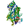
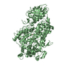
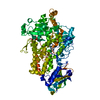
| Entry | Database: PDB / ID: 1n8q | ||||||
|---|---|---|---|---|---|---|---|
| Title | LIPOXYGENASE IN COMPLEX WITH PROTOCATECHUIC ACID | ||||||
 Components Components | lipoxygenase-3 | ||||||
 Keywords Keywords | OXIDOREDUCTASE / lipoxygenase / iron / protocatechuic acid / 3 / 4-dihydroxybenzoic acid / lox complex / quercetin | ||||||
| Function / homology |  Function and homology information Function and homology informationlinoleate 9S-lipoxygenase / linoleate 9S-lipoxygenase activity / oxylipin biosynthetic process / lipid oxidation / oxidoreductase activity, acting on single donors with incorporation of molecular oxygen, incorporation of two atoms of oxygen / fatty acid biosynthetic process / iron ion binding / cytoplasm Similarity search - Function | ||||||
| Biological species |  | ||||||
| Method |  X-RAY DIFFRACTION / MOLECULAR REPLACEMENT / Resolution: 2.1 Å X-RAY DIFFRACTION / MOLECULAR REPLACEMENT / Resolution: 2.1 Å | ||||||
 Authors Authors | Borbulevych, O.Y. / Jankun, J. / Selman, S.H. / Skrzypczak-Jankun, E. | ||||||
 Citation Citation | Journal: PROTEINS: STRUCT.,FUNCT.,GENET. / Year: 2004 Title: Lipoxygenase interactions with natural flavonoid, quercetin, reveal a complex with protocatechuic acid in its X-ray structure at 2.1 A resolution. Authors: Borbulevych, O.Y. / Jankun, J. / Selman, S.H. / Skrzypczak-Jankun, E. #1: Journal: Biochemistry / Year: 1998Title: Structural and thermochemical characterization of lipoxygenase-catechol complexes Authors: Pham, C.H. / Jankun, J. / Skrzypczak-Jankun, E. / Flowers II, R.A. / Funk Jr., M.O. #2: Journal: Int.J.Mol.Med. / Year: 2000Title: Curcumin inhibits lipoxygenase by binding to its central cavity: Theoretical and X-ray evidence Authors: Skrzypczak-Jankun, E. / McCabe, N.P. / Selman, S.H. / Jankun, J. | ||||||
| History |
|
- Structure visualization
| Structure viewer | Molecule: MolmilJmol/JSmol |
|---|
- Downloads & links
-Download
| PDBx/mmCIF format | 1n8q.cif.gz | 191.6 KB | Display | PDBx/mmCIF format |
|---|---|---|---|---|
| PDB format | pdb1n8q.ent.gz | 150 KB | Display | PDB format |
| PDBx/mmJSON format | 1n8q.json.gz | Tree view | PDBx/mmJSON format | |
| Others |  Other downloads Other downloads |
-Validation report
| Arichive directory | https://data.pdbj.org/pub/pdb/validation_reports/n8/1n8qftp://data.pdbj.org/pub/pdb/validation_reports/n8/1n8q | HTTPS FTP |
|---|
-Related structure data
| Related structure data |  1ik3S S: Starting model for refinement |
|---|---|
| Similar structure data |


-Links
 PDBj
PDBj


- Assembly
| Deposited unit | 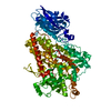
| ||||||||
|---|---|---|---|---|---|---|---|---|---|
| 1 |
| ||||||||
| Unit cell |
| ||||||||
| Components on special symmetry positions |
|
-Components
| #1: Protein | Mass: 96919.000 Da / Num. of mol.: 1 / Source method: isolated from a natural source / Source: (natural) |
|---|---|
| #2: Chemical | ChemComp-FE2 /   Mass: 55.845 Da / Num. of mol.: 1 / Source method: obtained synthetically / Formula: Fe Mass: 55.845 Da / Num. of mol.: 1 / Source method: obtained synthetically / Formula: Fe |
| #3: Chemical | ChemComp-DHB / 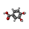  Mass: 154.120 Da / Num. of mol.: 1 / Source method: obtained synthetically / Formula: C7H6O4 Mass: 154.120 Da / Num. of mol.: 1 / Source method: obtained synthetically / Formula: C7H6O4 |
| #4: Water | ChemComp-HOH /  Mass: 18.015 Da / Num. of mol.: 463 / Source method: isolated from a natural source / Formula: H2O Mass: 18.015 Da / Num. of mol.: 463 / Source method: isolated from a natural source / Formula: H2O |
-Experimental details
-Experiment
| Experiment | Method: X-RAY DIFFRACTION / Number of used crystals: 3 |
|---|
- Sample preparation
| Crystal | Density Matthews: 2.34 Å3/Da / Density % sol: 46.95 % | |||||||||||||||||||||||||||||||||||||||||||||||||
|---|---|---|---|---|---|---|---|---|---|---|---|---|---|---|---|---|---|---|---|---|---|---|---|---|---|---|---|---|---|---|---|---|---|---|---|---|---|---|---|---|---|---|---|---|---|---|---|---|---|---|
| Crystal grow | Temperature: 295 K / Method: vapor diffusion, sitting drop / pH: 5.3 Details: PEG 8000, citrate-phosphate buffer, tris HCl, sodium azide, pH 5.3, VAPOR DIFFUSION, SITTING DROP, temperature 295K | |||||||||||||||||||||||||||||||||||||||||||||||||
| Crystal grow | *PLUS Temperature: 23 ℃ / pH: 7 / Method: vapor diffusion, sitting drop / Details: Skrzypczak-Jankun, E., (1997) Proteins, 29, 15. | |||||||||||||||||||||||||||||||||||||||||||||||||
| Components of the solutions | *PLUS
|
-Data collection
| Diffraction | Mean temperature: 295 K |
|---|---|
| Diffraction source | Source: ROTATING ANODE / Type: RIGAKU RU200 / Wavelength: 1.5418 Å |
| Detector | Type: RIGAKU RAXIS IV / Detector: IMAGE PLATE / Date: Jan 15, 2002 / Details: Focusing mirrors |
| Radiation | Protocol: SINGLE WAVELENGTH / Monochromatic (M) / Laue (L): M / Scattering type: x-ray |
| Radiation wavelength | Wavelength: 1.5418 Å / Relative weight: 1 |
| Reflection | Resolution: 2.1→50 Å / Num. all: 54294 / Num. obs: 52285 / % possible obs: 0.963 % / Observed criterion σ(F): 0 / Observed criterion σ(I): 0 / Redundancy: 2 % / Biso Wilson estimate: 37.7 Å2 / Rmerge(I) obs: 0.086 / Net I/σ(I): 12 |
| Reflection shell | Resolution: 2.1→2.18 Å / Redundancy: 2 % / Rmerge(I) obs: 0.471 / Mean I/σ(I) obs: 1.36 / Num. unique all: 4668 / % possible all: 86.2 |
| Reflection | *PLUS Highest resolution: 2.1 Å / Lowest resolution: 50 Å / Num. obs: 52281 / Rmerge(I) obs: 0.07 |
| Reflection shell | *PLUS % possible obs: 86.2 % / Rmerge(I) obs: 0.45 |
- Processing
| Software |
| ||||||||||||||||||||||||||||||||||||||||||||||||||||||||||||||||||||||
|---|---|---|---|---|---|---|---|---|---|---|---|---|---|---|---|---|---|---|---|---|---|---|---|---|---|---|---|---|---|---|---|---|---|---|---|---|---|---|---|---|---|---|---|---|---|---|---|---|---|---|---|---|---|---|---|---|---|---|---|---|---|---|---|---|---|---|---|---|---|---|---|
| Refinement | Method to determine structure: MOLECULAR REPLACEMENT Starting model: PDB ENTRY 1IK3 Resolution: 2.1→50 Å / Cor.coef. Fo:Fc: 0.956 / Cor.coef. Fo:Fc free: 0.927 / SU B: 5.608 / SU ML: 0.147 / TLS residual ADP flag: LIKELY RESIDUAL / Isotropic thermal model: isotropic thermal model / Cross valid method: THROUGHOUT / σ(F): 0 / ESU R: 0.243 / ESU R Free: 0.203 / Stereochemistry target values: MAXIMUM LIKELIHOOD
| ||||||||||||||||||||||||||||||||||||||||||||||||||||||||||||||||||||||
| Solvent computation | Ion probe radii: 0.8 Å / Shrinkage radii: 0.8 Å / VDW probe radii: 1.4 Å / Solvent model: BABINET MODEL WITH MASK | ||||||||||||||||||||||||||||||||||||||||||||||||||||||||||||||||||||||
| Displacement parameters | Biso mean: 31.753 Å2
| ||||||||||||||||||||||||||||||||||||||||||||||||||||||||||||||||||||||
| Refine analyze | Luzzati coordinate error obs: 0.304 Å / Luzzati d res low obs: 8 Å | ||||||||||||||||||||||||||||||||||||||||||||||||||||||||||||||||||||||
| Refinement step | Cycle: LAST / Resolution: 2.1→50 Å
| ||||||||||||||||||||||||||||||||||||||||||||||||||||||||||||||||||||||
| Refine LS restraints |
| ||||||||||||||||||||||||||||||||||||||||||||||||||||||||||||||||||||||
| LS refinement shell | Resolution: 2.1→2.154 Å / Total num. of bins used: 20
| ||||||||||||||||||||||||||||||||||||||||||||||||||||||||||||||||||||||
| Refinement TLS params. | Method: refined / Origin x: 27.3399 Å / Origin y: 0.503 Å / Origin z: 15.2134 Å
| ||||||||||||||||||||||||||||||||||||||||||||||||||||||||||||||||||||||
| Refinement | *PLUS Highest resolution: 2.1 Å / Lowest resolution: 50 Å | ||||||||||||||||||||||||||||||||||||||||||||||||||||||||||||||||||||||
| Solvent computation | *PLUS | ||||||||||||||||||||||||||||||||||||||||||||||||||||||||||||||||||||||
| Displacement parameters | *PLUS | ||||||||||||||||||||||||||||||||||||||||||||||||||||||||||||||||||||||
| Refine LS restraints | *PLUS
|
