 Movie
Movie Controller


+ Open data

- Basic information
| Entry | Database: PDB / ID: 1n6h | ||||||
|---|---|---|---|---|---|---|---|
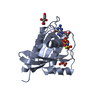
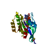
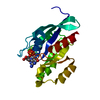
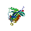
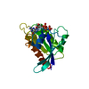
| Title | Crystal Structure of Human Rab5a | ||||||
 Components Components | Ras-related protein Rab-5A | ||||||
 Keywords Keywords | PROTEIN TRANSPORT / Rab / GTPase | ||||||
| Function / homology |  Function and homology information Function and homology informationregulation of endosome size / cytoplasmic side of early endosome membrane / protein localization to early endosome / amyloid-beta clearance by transcytosis / synaptic vesicle recycling / plasma membrane to endosome transport / host-mediated perturbation of viral process / regulation of filopodium assembly / early endosome to late endosome transport / RAB geranylgeranylation ...regulation of endosome size / cytoplasmic side of early endosome membrane / protein localization to early endosome / amyloid-beta clearance by transcytosis / synaptic vesicle recycling / plasma membrane to endosome transport / host-mediated perturbation of viral process / regulation of filopodium assembly / early endosome to late endosome transport / RAB geranylgeranylation / regulation of autophagosome assembly / RAB GEFs exchange GTP for GDP on RABs / early phagosome / TBC/RABGAPs / endosomal transport / regulation of synaptic vesicle exocytosis / Synthesis of PIPs at the plasma membrane / positive regulation of exocytosis / Respiratory syncytial virus (RSV) attachment and entry / endocytic vesicle / canonical Wnt signaling pathway / phagocytosis / phagocytic vesicle / ruffle / somatodendritic compartment / endomembrane system / Prevention of phagosomal-lysosomal fusion / axon terminus / small monomeric GTPase / clathrin-coated endocytic vesicle membrane / intracellular protein transport / receptor internalization / regulation of long-term neuronal synaptic plasticity / phagocytic vesicle membrane / endocytosis / terminal bouton / synaptic vesicle / melanosome / GDP binding / actin cytoskeleton / synaptic vesicle membrane / Clathrin-mediated endocytosis / Factors involved in megakaryocyte development and platelet production / G protein activity / early endosome membrane / early endosome / endosome / endosome membrane / membrane raft / axon / neuronal cell body / GTPase activity / dendrite / GTP binding / extracellular exosome / nucleoplasm / plasma membrane / cytoplasm / cytosol Similarity search - Function | ||||||
| Biological species |  Homo sapiens (human) Homo sapiens (human) | ||||||
| Method |  X-RAY DIFFRACTION / FOURIER SYNTHESIS / Resolution: 1.51 Å X-RAY DIFFRACTION / FOURIER SYNTHESIS / Resolution: 1.51 Å | ||||||
 Authors Authors | Zhu, G. / Liu, J. / Terzyan, S. / Zhai, P. / Li, G. / Zhang, X.C. | ||||||
 Citation Citation | Journal: J.Biol.Chem. / Year: 2003 Title: High Resolution Crystal Structures of Human Rab5a and Five Mutants with Substitutions in the Catalytically Important Phosphate-Binding Loop Authors: Zhu, G. / Liu, J. / Terzyan, S. / Zhai, P. / Li, G. / Zhang, X.C. | ||||||
| History |
|
- Structure visualization
| Structure viewer | Molecule: MolmilJmol/JSmol |
|---|
- Downloads & links
-Download
| PDBx/mmCIF format | 1n6h.cif.gz | 56 KB | Display | PDBx/mmCIF format |
|---|---|---|---|---|
| PDB format | pdb1n6h.ent.gz | 39 KB | Display | PDB format |
| PDBx/mmJSON format | 1n6h.json.gz | Tree view | PDBx/mmJSON format | |
| Others |  Other downloads Other downloads |
-Validation report
| Arichive directory | https://data.pdbj.org/pub/pdb/validation_reports/n6/1n6hftp://data.pdbj.org/pub/pdb/validation_reports/n6/1n6h | HTTPS FTP |
|---|
-Related structure data
| Related structure data | 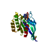 1n6iC 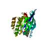 1n6kC 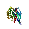 1n6lC 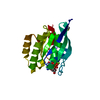 1n6nC 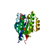 1n6oC 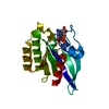 1n6pC  1n6rC 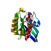 1huqS C: citing same article ( S: Starting model for refinement |
|---|---|
| Similar structure data |


-Links
 PDBj
PDBj


- Assembly
| Deposited unit | 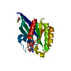
| ||||||||
|---|---|---|---|---|---|---|---|---|---|
| 1 |
| ||||||||
| Unit cell |
|
-Components
| #1: Protein | Mass: 18987.572 Da / Num. of mol.: 1 / Fragment: GTPASE DOMAIN Source method: isolated from a genetically manipulated source Source: (gene. exp.) Homo sapiens (human) / Plasmid: pET11a / Species (production host): Escherichia coli / Production host:  |
|---|---|
| #2: Chemical | ChemComp-MG /   Mass: 24.305 Da / Num. of mol.: 1 / Source method: obtained synthetically / Formula: Mg Mass: 24.305 Da / Num. of mol.: 1 / Source method: obtained synthetically / Formula: Mg |
| #3: Chemical | ChemComp-GNP /   Mass: 522.196 Da / Num. of mol.: 1 / Source method: obtained synthetically / Formula: C10H17N6O13P3 Mass: 522.196 Da / Num. of mol.: 1 / Source method: obtained synthetically / Formula: C10H17N6O13P3Comment: GppNHp, GMPPNP, energy-carrying molecule analogue*YM |
| #4: Chemical | ChemComp-BME /   Mass: 78.133 Da / Num. of mol.: 1 / Source method: obtained synthetically / Formula: C2H6OS Mass: 78.133 Da / Num. of mol.: 1 / Source method: obtained synthetically / Formula: C2H6OS |
| #5: Water | ChemComp-HOH /  Mass: 18.015 Da / Num. of mol.: 263 / Source method: isolated from a natural source / Formula: H2O Mass: 18.015 Da / Num. of mol.: 263 / Source method: isolated from a natural source / Formula: H2O |
| Has protein modification | N |
-Experimental details
-Experiment
| Experiment | Method: X-RAY DIFFRACTION / Number of used crystals: 1 |
|---|
- Sample preparation
| Crystal | Density Matthews: 2 Å3/Da / Density % sol: 37.5 % | |||||||||||||||||||||||||||||||||||||||||||||||||||||||||||||||
|---|---|---|---|---|---|---|---|---|---|---|---|---|---|---|---|---|---|---|---|---|---|---|---|---|---|---|---|---|---|---|---|---|---|---|---|---|---|---|---|---|---|---|---|---|---|---|---|---|---|---|---|---|---|---|---|---|---|---|---|---|---|---|---|---|
| Crystal grow | Temperature: 277 K / Method: vapor diffusion, hanging drop / pH: 6 Details: PEG 6000, sodium chloride, MES, pH 6.0, VAPOR DIFFUSION, HANGING DROP, temperature 277K | |||||||||||||||||||||||||||||||||||||||||||||||||||||||||||||||
| Crystal grow | *PLUS pH: 8 | |||||||||||||||||||||||||||||||||||||||||||||||||||||||||||||||
| Components of the solutions | *PLUS
|
-Data collection
| Diffraction | Mean temperature: 100 K |
|---|---|
| Diffraction source | Source: ROTATING ANODE / Type: RIGAKU / Wavelength: 1.5418 Å |
| Detector | Type: MARRESEARCH / Detector: IMAGE PLATE / Date: Feb 23, 2002 |
| Radiation | Monochromator: OSMIC OPTICS / Protocol: SINGLE WAVELENGTH / Monochromatic (M) / Laue (L): M / Scattering type: x-ray |
| Radiation wavelength | Wavelength: 1.5418 Å / Relative weight: 1 |
| Reflection | Resolution: 1.51→20 Å / Num. all: 24442 / Num. obs: 24259 / % possible obs: 98.9 % / Observed criterion σ(F): 0 / Observed criterion σ(I): 0 / Redundancy: 5.5 % / Biso Wilson estimate: 18.8 Å2 / Rmerge(I) obs: 0.035 / Net I/σ(I): 36.3 |
| Reflection shell | Resolution: 1.51→1.56 Å / Rmerge(I) obs: 0.14 / Mean I/σ(I) obs: 5.2 / % possible all: 97.7 |
| Reflection | *PLUS Lowest resolution: 20 Å / Num. obs: 24442 / % possible obs: 99.5 % / Num. measured all: 134293 |
| Reflection shell | *PLUS % possible obs: 99.3 % |
- Processing
| Software |
| ||||||||||||||||||||||||||||||||||||
|---|---|---|---|---|---|---|---|---|---|---|---|---|---|---|---|---|---|---|---|---|---|---|---|---|---|---|---|---|---|---|---|---|---|---|---|---|---|
| Refinement | Method to determine structure: FOURIER SYNTHESIS Starting model: PDB ENTRY 1HUQ Resolution: 1.51→20 Å / Cross valid method: THROUGHOUT / σ(F): 0 / Stereochemistry target values: Engh & Huber
| ||||||||||||||||||||||||||||||||||||
| Displacement parameters |
| ||||||||||||||||||||||||||||||||||||
| Refine analyze |
| ||||||||||||||||||||||||||||||||||||
| Refinement step | Cycle: LAST / Resolution: 1.51→20 Å
| ||||||||||||||||||||||||||||||||||||
| Refine LS restraints |
| ||||||||||||||||||||||||||||||||||||
| Xplor file |
| ||||||||||||||||||||||||||||||||||||
| Refinement | *PLUS Lowest resolution: 20 Å / Rfactor Rfree: 0.189 / Rfactor Rwork: 0.176 | ||||||||||||||||||||||||||||||||||||
| Solvent computation | *PLUS | ||||||||||||||||||||||||||||||||||||
| Displacement parameters | *PLUS | ||||||||||||||||||||||||||||||||||||
| Refine LS restraints | *PLUS
|
