 Movie
Movie Controller

[English] 日本語
 Yorodumi
- PDB-1n5s: Crystal structure of a Monooxygenase from the gene ActVA-Orf6 of ... -
+ Open data

- Basic information
| Entry | Database: PDB / ID: 1n5s | ||||||
|---|---|---|---|---|---|---|---|
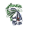
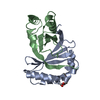
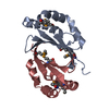
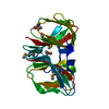
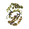
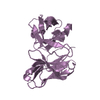
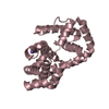
| Title | Crystal structure of a Monooxygenase from the gene ActVA-Orf6 of Streptomyces coelicolor in complex with the ligand Acetyl Dithranol | ||||||
 Components Components | ActVA-Orf6 monooxygenase | ||||||
 Keywords Keywords | OXIDOREDUCTASE / monooxygenase / aromatic polyketides / actinorhodin / dihydrokalafungin / acetyl dithranol / streptomyces coelicolor | ||||||
| Function / homology |  Function and homology information Function and homology informationABM domain profile. / Antibiotic biosynthesis monooxygenase / Antibiotic biosynthesis monooxygenase domain / Alpha-Beta Plaits - #100 / Dimeric alpha-beta barrel / Alpha-Beta Plaits / 2-Layer Sandwich / Alpha Beta Similarity search - Domain/homology | ||||||
| Biological species |  Streptomyces coelicolor (bacteria) Streptomyces coelicolor (bacteria) | ||||||
| Method |  X-RAY DIFFRACTION / SYNCHROTRON / fourier difference / Resolution: 1.7 Å X-RAY DIFFRACTION / SYNCHROTRON / fourier difference / Resolution: 1.7 Å | ||||||
 Authors Authors | Sciara, G. / Kendrew, S.G. / Miele, A.E. / Marsh, N.G. / Federici, L. / Malatesta, F. / Schimperna, G. / Savino, C. / Vallone, B. | ||||||
 Citation Citation | Journal: Embo J. / Year: 2003 Title: The structure of ActVA-Orf6, a novel type of monooxygenase involved in actinorhodin biosynthesis Authors: Sciara, G. / Kendrew, S.G. / Miele, A.E. / Marsh, N.G. / Federici, L. / Malatesta, F. / Schimperna, G. / Savino, C. / Vallone, B. #1: Journal: Acta Crystallogr.,Sect.D / Year: 2000Title: Crystallization and preliminary X-ray diffraction studies of a monooxygenase from Streptomyces coelicolor A3(2) involved in the biosynthesis of the polyketide actinorhodin Authors: Kendrew, S.G. / Federici, L. / Savino, C. / Miele, A.E. / Marsh, N.G. / Vallone, B. #2: Journal: J.Bacteriol. / Year: 1997Title: Identification of a monooxygenase from Streptomyces coelicolor A3 (2) involved in the biosynthesis of actinorhodin: purification and characterization of the recombinant enzyme Authors: Kendrew, S.G. / Hopwood, D.A. / Marsh, N.G. | ||||||
| History |
|
- Structure visualization
| Structure viewer | Molecule: MolmilJmol/JSmol |
|---|
- Downloads & links
-Download
| PDBx/mmCIF format | 1n5s.cif.gz | 59.1 KB | Display | PDBx/mmCIF format |
|---|---|---|---|---|
| PDB format | pdb1n5s.ent.gz | 43.5 KB | Display | PDB format |
| PDBx/mmJSON format | 1n5s.json.gz | Tree view | PDBx/mmJSON format | |
| Others |  Other downloads Other downloads |
-Validation report
| Arichive directory | https://data.pdbj.org/pub/pdb/validation_reports/n5/1n5sftp://data.pdbj.org/pub/pdb/validation_reports/n5/1n5s | HTTPS FTP |
|---|
-Related structure data
| Related structure data |  1lq9SC  1n5qC 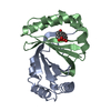 1n5tC  1n5vC S: Starting model for refinement C: citing same article ( |
|---|---|
| Similar structure data |


-Links
 PDBj
PDBj- Assembly
| Deposited unit | 
| ||||||||
|---|---|---|---|---|---|---|---|---|---|
| 1 |
| ||||||||
| Unit cell |
| ||||||||
| Details | The biological unit is a dimer in the asymmetric unit |
-Components
| #1: Protein | Mass: 11978.420 Da / Num. of mol.: 2 Source method: isolated from a genetically manipulated source Source: (gene. exp.) Streptomyces coelicolor (bacteria) / Strain: A3(2) / Gene: ActVA-Orf6 / Plasmid: pT7-7 / Production host: #2: Chemical | ChemComp-1PG / |   Mass: 252.305 Da / Num. of mol.: 1 / Source method: obtained synthetically / Formula: C11H24O6 Mass: 252.305 Da / Num. of mol.: 1 / Source method: obtained synthetically / Formula: C11H24O6#3: Chemical | ChemComp-ADL / ( |   Mass: 284.263 Da / Num. of mol.: 1 / Source method: obtained synthetically / Formula: C16H12O5 Mass: 284.263 Da / Num. of mol.: 1 / Source method: obtained synthetically / Formula: C16H12O5#4: Water | ChemComp-HOH / |  Mass: 18.015 Da / Num. of mol.: 158 / Source method: isolated from a natural source / Formula: H2O Mass: 18.015 Da / Num. of mol.: 158 / Source method: isolated from a natural source / Formula: H2O |
|---|
-Experimental details
-Experiment
| Experiment | Method: X-RAY DIFFRACTION / Number of used crystals: 1 |
|---|
- Sample preparation
| Crystal | Density Matthews: 1.9 Å3/Da / Density % sol: 34.58 % | ||||||||||||||||||||||||||||||
|---|---|---|---|---|---|---|---|---|---|---|---|---|---|---|---|---|---|---|---|---|---|---|---|---|---|---|---|---|---|---|---|
| Crystal grow | Temperature: 293 K / Method: vapor diffusion + soaking / pH: 7 Details: ammonium sulfate, PEG 200, Tris or Hepes buffer , pH 7.0, vapour diffusion + soaking, temperature 293K | ||||||||||||||||||||||||||||||
| Crystal grow | *PLUS Method: vapor diffusion, hanging drop | ||||||||||||||||||||||||||||||
| Components of the solutions | *PLUS
|
-Data collection
| Diffraction | Mean temperature: 100 K |
|---|---|
| Diffraction source | Source: SYNCHROTRON / Site: ELETTRA  / Beamline: 5.2R / Wavelength: 1 Å / Beamline: 5.2R / Wavelength: 1 Å |
| Detector | Type: MARRESEARCH / Detector: CCD / Date: Mar 25, 2001 / Details: Three-segment Pt-coated toroidal mirror |
| Radiation | Monochromator: Double crystal (Si111,Si220) / Protocol: SINGLE WAVELENGTH / Monochromatic (M) / Laue (L): M / Scattering type: x-ray |
| Radiation wavelength | Wavelength: 1 Å / Relative weight: 1 |
| Reflection | Resolution: 1.7→20 Å / Num. all: 22894 / Num. obs: 22894 / % possible obs: 100 % / Redundancy: 10 % / Biso Wilson estimate: 20.29 Å2 / Rmerge(I) obs: 0.025 / Net I/σ(I): 99.6 |
| Reflection shell | Resolution: 1.7→1.73 Å / Rmerge(I) obs: 0.098 / Mean I/σ(I) obs: 18.3 / Num. unique all: 1135 / % possible all: 99.8 |
| Reflection | *PLUS Num. measured all: 228029 |
| Reflection shell | *PLUS % possible obs: 99.8 % |
- Processing
| Software |
| |||||||||||||||||||||||||
|---|---|---|---|---|---|---|---|---|---|---|---|---|---|---|---|---|---|---|---|---|---|---|---|---|---|---|
| Refinement | Method to determine structure: fourier difference Starting model: PDB ENTRY 1LQ9 Resolution: 1.7→20 Å / Stereochemistry target values: Engh & Huber
| |||||||||||||||||||||||||
| Refinement step | Cycle: LAST / Resolution: 1.7→20 Å
| |||||||||||||||||||||||||
| Refinement | *PLUS % reflection Rfree: 5 % | |||||||||||||||||||||||||
| Solvent computation | *PLUS | |||||||||||||||||||||||||
| Displacement parameters | *PLUS | |||||||||||||||||||||||||
| Refine LS restraints | *PLUS
|
