 Movie
Movie Controller

[English] 日本語
 Yorodumi
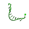
- PDB-1n5c: Crystal Structure Analysis of the B-DNA Dodecamer CGCGAATT(ethenoC)GCG -
+ Open data

- Basic information
| Entry | Database: PDB / ID: 1n5c | ||||||||||||||||||
|---|---|---|---|---|---|---|---|---|---|---|---|---|---|---|---|---|---|---|---|
| Title | Crystal Structure Analysis of the B-DNA Dodecamer CGCGAATT(ethenoC)GCG | ||||||||||||||||||
 Components Components | 5'-D(* Keywords KeywordsDNA / B form double helix / 3 / N4-etheno-2'-cytidine modification opposite G | Function / homology | DNA / DNA (> 10) |  Function and homology information Function and homology informationMethod |  X-RAY DIFFRACTION / SYNCHROTRON / ISOMORPHOUS REPLACEMENT / Resolution: 1.79 Å X-RAY DIFFRACTION / SYNCHROTRON / ISOMORPHOUS REPLACEMENT / Resolution: 1.79 Å  Authors AuthorsFreisinger, E. / Fernandes, A. / Grollman, A.P. / Kisker, C.F. |  CitationJournal: J.Mol.Biol. / Year: 2003 CitationJournal: J.Mol.Biol. / Year: 2003Title: Crystallographic Characterization of an Exocyclic DNA Adduct: 3,N4-etheno-2'-deoxycytidine in the Dodecamer 5'-CGCGAATT(ethenoC)GCG-3' Authors: Freisinger, E. / Fernandes, A. / Grollman, A.P. / Kisker, C.F. History |
|
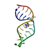
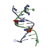
- Structure visualization
| Structure viewer | Molecule: MolmilJmol/JSmol |
|---|
- Downloads & links
-Download
| PDBx/mmCIF format | 1n5c.cif.gz | 19.3 KB | Display | PDBx/mmCIF format |
|---|---|---|---|---|
| PDB format | pdb1n5c.ent.gz | 11.4 KB | Display | PDB format |
| PDBx/mmJSON format | 1n5c.json.gz | Tree view | PDBx/mmJSON format | |
| Others |  Other downloads Other downloads |
-Validation report
| Arichive directory | https://data.pdbj.org/pub/pdb/validation_reports/n5/1n5cftp://data.pdbj.org/pub/pdb/validation_reports/n5/1n5c | HTTPS FTP |
|---|
-Related structure data
| Similar structure data |
|---|

-Links
 PDBj
PDBj


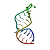
- Assembly
| Deposited unit | 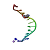
| ||||||||||||||||||
|---|---|---|---|---|---|---|---|---|---|---|---|---|---|---|---|---|---|---|---|
| 1 | 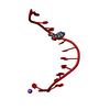
| ||||||||||||||||||
| Unit cell |
| ||||||||||||||||||
| Components on special symmetry positions |
| ||||||||||||||||||
| Details | The second part of the biological assembly/double helix is generated by the two fold axis: -x+y+1, y, -z+3/2. |
-Components
| #1: DNA chain | Mass: 3687.414 Da / Num. of mol.: 1 / Source method: obtained synthetically |
|---|---|
| #2: Chemical | ChemComp-NA /   Mass: 22.990 Da / Num. of mol.: 1 / Source method: obtained synthetically / Formula: Na Mass: 22.990 Da / Num. of mol.: 1 / Source method: obtained synthetically / Formula: Na |
| #3: Water | ChemComp-HOH /  Mass: 18.015 Da / Num. of mol.: 73 / Source method: isolated from a natural source / Formula: H2O Mass: 18.015 Da / Num. of mol.: 73 / Source method: isolated from a natural source / Formula: H2O |
-Experimental details
-Experiment
| Experiment | Method: X-RAY DIFFRACTION / Number of used crystals: 1 |
|---|
- Sample preparation
| Crystal | Density Matthews: 2.34 Å3/Da / Density % sol: 47.11 % | ||||||||||||||||||||||||||||||||||||||||||
|---|---|---|---|---|---|---|---|---|---|---|---|---|---|---|---|---|---|---|---|---|---|---|---|---|---|---|---|---|---|---|---|---|---|---|---|---|---|---|---|---|---|---|---|
| Crystal grow | Temperature: 277 K / pH: 7 Details: Drop: MPD, potassium chloride, spermine tetrahydrochloride, cacodylate; Reservoir: MPD, pH 7.0, VAPOR DIFFUSION, HANGING DROP, temperature 277K, pH 7.00 | ||||||||||||||||||||||||||||||||||||||||||
| Components of the solutions |
| ||||||||||||||||||||||||||||||||||||||||||
| Crystal grow | *PLUS Temperature: 4 ℃ / pH: 7 / Method: vapor diffusion, hanging drop | ||||||||||||||||||||||||||||||||||||||||||
| Components of the solutions | *PLUS
|
-Data collection
| Diffraction | Mean temperature: 100 K |
|---|---|
| Diffraction source | Source: SYNCHROTRON / Site: NSLS  / Beamline: X26C / Wavelength: 1.1 / Beamline: X26C / Wavelength: 1.1 |
| Detector | Type: ADSC QUANTUM 4 / Detector: CCD / Date: Jun 13, 2000 |
| Radiation | Monochromator: MIRROR / Protocol: SINGLE WAVELENGTH / Monochromatic (M) / Laue (L): M / Scattering type: x-ray |
| Radiation wavelength | Wavelength: 1.1 Å / Relative weight: 1 |
| Reflection | Resolution: 1.79→50 Å / Num. obs: 4113 / % possible obs: 99.4 % / Observed criterion σ(I): 5 / Redundancy: 4 % / Rsym value: 0.061 / Net I/σ(I): 22 |
| Reflection shell | Resolution: 1.79→1.85 Å / Redundancy: 4 % / Mean I/σ(I) obs: 2 / Rsym value: 0.187 / % possible all: 100 |
| Reflection | *PLUS Highest resolution: 1.79 Å / Lowest resolution: 50 Å / Rmerge(I) obs: 0.061 |
- Processing
| Software |
| |||||||||||||||||||||||||||||||||
|---|---|---|---|---|---|---|---|---|---|---|---|---|---|---|---|---|---|---|---|---|---|---|---|---|---|---|---|---|---|---|---|---|---|---|
| Refinement | Method to determine structure: ISOMORPHOUS REPLACEMENT Starting model: NDB ENTRY BD0032 Resolution: 1.79→20 Å / Num. parameters: 1327 / Num. restraintsaints: 1445 / Cross valid method: THROUGHOUT / σ(F): 0 / Stereochemistry target values: ENGH & HUBER
| |||||||||||||||||||||||||||||||||
| Solvent computation | Solvent model: BABINET'S PRINCIPLE | |||||||||||||||||||||||||||||||||
| Refine analyze | Occupancy sum hydrogen: 137 / Occupancy sum non hydrogen: 303.75 | |||||||||||||||||||||||||||||||||
| Refinement step | Cycle: LAST / Resolution: 1.79→20 Å
| |||||||||||||||||||||||||||||||||
| Refine LS restraints |
| |||||||||||||||||||||||||||||||||
| Software | *PLUS Name: SHELXL / Version: 97 / Classification: refinement | |||||||||||||||||||||||||||||||||
| Refinement | *PLUS Rfactor obs: 0.1612 / Rfactor Rfree: 0.2193 / Rfactor Rwork: 0.061 | |||||||||||||||||||||||||||||||||
| Solvent computation | *PLUS | |||||||||||||||||||||||||||||||||
| Displacement parameters | *PLUS | |||||||||||||||||||||||||||||||||
| Refine LS restraints | *PLUS
|
