 Movie
Movie Controller

[English] 日本語
 Yorodumi
- PDB-1n1c: Crystal Structure Of The Dimeric TorD Chaperone From Shewanella M... -
+ Open data

- Basic information
| Entry | Database: PDB / ID: 1n1c | ||||||
|---|---|---|---|---|---|---|---|
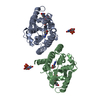
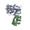
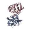
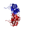
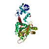
| Title | Crystal Structure Of The Dimeric TorD Chaperone From Shewanella Massilia | ||||||
 Components Components | TorA specific chaperone | ||||||
 Keywords Keywords | CHAPERONE / TORD / 3D-domain swapping | ||||||
| Function / homology |  Function and homology information Function and homology information | ||||||
| Biological species |  Shewanella massilia (bacteria) Shewanella massilia (bacteria) | ||||||
| Method |  X-RAY DIFFRACTION / SYNCHROTRON / MAD / Resolution: 2.4 Å X-RAY DIFFRACTION / SYNCHROTRON / MAD / Resolution: 2.4 Å | ||||||
 Authors Authors | Tranier, S. / Iobbi-Nivol, C. / Mortier-Barriere, I. / Birck, C. / Mejean, V. / Samama, J.-P. | ||||||
 Citation Citation | Journal: Structure / Year: 2003 Title: A Novel Protein Fold and Extreme Domain Swapping in the Dimeric TorD Chaperone from Shewanella massilia Authors: Tranier, S. / Iobbi-Nivol, C. / Birck, C. / Ilbert, M. / Mortier-Barriere, I. / Mejean, V. / Samama, J.P. #1: Journal: Protein Sci. / Year: 2002Title: Characterization And Multiple Molecular Forms Of TorD From Shewanella Massilia, The Putative Chaperone Of The Molybdoenzyme TorA Authors: Tranier, S. / Mortier-Barriere, I. / Ilbert, M. / Birck, C. / Iobbi-Nivol, C. / Mejean, V. / Samama, J.-P. #2: Journal: J.Biol.Chem. / Year: 1998Title: TorD, a cytoplasmic chaperone that interacts with the unfolded trimethylamine N-oxide reductase enzyme (TorA) in Escherichia coli Authors: Pommier, J. / Mejean, V. / Giordano, G. / Iobbi-Nivol, C. | ||||||
| History |
|
- Structure visualization
| Structure viewer | Molecule: MolmilJmol/JSmol |
|---|
- Downloads & links
-Download
| PDBx/mmCIF format | 1n1c.cif.gz | 93.1 KB | Display | PDBx/mmCIF format |
|---|---|---|---|---|
| PDB format | pdb1n1c.ent.gz | 71.6 KB | Display | PDB format |
| PDBx/mmJSON format | 1n1c.json.gz | Tree view | PDBx/mmJSON format | |
| Others |  Other downloads Other downloads |
-Validation report
| Arichive directory | https://data.pdbj.org/pub/pdb/validation_reports/n1/1n1cftp://data.pdbj.org/pub/pdb/validation_reports/n1/1n1c | HTTPS FTP |
|---|
-Related structure data
| Similar structure data |
|---|


-Links
 PDBj
PDBj


- Assembly
| Deposited unit | 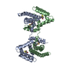
| ||||||||
|---|---|---|---|---|---|---|---|---|---|
| 1 |
| ||||||||
| Unit cell |
|
-Components
| #1: Protein | Mass: 24654.322 Da / Num. of mol.: 2 / Mutation: H8P/F147C/F163V Source method: isolated from a genetically manipulated source Source: (gene. exp.) Shewanella massilia (bacteria) / Gene: torD / Plasmid: pet-22b / Production host: #2: Chemical |   Mass: 154.251 Da / Num. of mol.: 2 / Source method: obtained synthetically / Formula: C4H10O2S2 Mass: 154.251 Da / Num. of mol.: 2 / Source method: obtained synthetically / Formula: C4H10O2S2#3: Water | ChemComp-HOH / |  Mass: 18.015 Da / Num. of mol.: 144 / Source method: isolated from a natural source / Formula: H2O Mass: 18.015 Da / Num. of mol.: 144 / Source method: isolated from a natural source / Formula: H2OHas protein modification | Y | |
|---|
-Experimental details
-Experiment
| Experiment | Method: X-RAY DIFFRACTION / Number of used crystals: 1 |
|---|
- Sample preparation
| Crystal | Density Matthews: 3.08 Å3/Da / Density % sol: 59.75 % | |||||||||||||||||||||||||||||||||||||||||||||||||
|---|---|---|---|---|---|---|---|---|---|---|---|---|---|---|---|---|---|---|---|---|---|---|---|---|---|---|---|---|---|---|---|---|---|---|---|---|---|---|---|---|---|---|---|---|---|---|---|---|---|---|
| Crystal grow | Temperature: 277 K / Method: vapor diffusion, hanging drop / pH: 6.4 Details: ammonium sulfate 1.6M, MES 100mM; Crystals were obtained by mixing 1 microl (1.2mg.ml-1) of protein solution with an equal volume of reservoir solution; Crystal Size (300x200x100 microM3), ...Details: ammonium sulfate 1.6M, MES 100mM; Crystals were obtained by mixing 1 microl (1.2mg.ml-1) of protein solution with an equal volume of reservoir solution; Crystal Size (300x200x100 microM3), pH 6.4, VAPOR DIFFUSION, HANGING DROP, temperature 277K | |||||||||||||||||||||||||||||||||||||||||||||||||
| Crystal grow | *PLUS Temperature: 4 ℃ / pH: 8 | |||||||||||||||||||||||||||||||||||||||||||||||||
| Components of the solutions | *PLUS
|
-Data collection
| Diffraction | Mean temperature: 100 K | ||||||||||||
|---|---|---|---|---|---|---|---|---|---|---|---|---|---|
| Diffraction source | Source: SYNCHROTRON / Site: EMBL/DESY, HAMBURG  / Beamline: BW7A / Wavelength: 0.97856, 0.95370, 0.97872 / Beamline: BW7A / Wavelength: 0.97856, 0.95370, 0.97872 | ||||||||||||
| Detector | Type: MARRESEARCH / Detector: CCD / Date: Aug 21, 2000 | ||||||||||||
| Radiation | Monochromator: DOUBLE CRYSTAL FOCUSSING MONOCHROMATOR / Protocol: MAD / Monochromatic (M) / Laue (L): M / Scattering type: x-ray | ||||||||||||
| Radiation wavelength |
| ||||||||||||
| Reflection | Resolution: 2.4→54.2 Å / Num. all: 23325 / Num. obs: 23239 / % possible obs: 98.7 % / Observed criterion σ(F): 0 / Observed criterion σ(I): 0 / Redundancy: 3.7 % / Biso Wilson estimate: 41.5 Å2 / Rmerge(I) obs: 0.033 / Net I/σ(I): 14 | ||||||||||||
| Reflection shell | Resolution: 2.4→2.53 Å / Redundancy: 3.7 % / Rmerge(I) obs: 0.177 / Mean I/σ(I) obs: 3.6 / Num. unique all: 3365 / % possible all: 98.9 | ||||||||||||
| Reflection | *PLUS Highest resolution: 2.4 Å / Lowest resolution: 54.23 Å / Num. measured all: 85570 | ||||||||||||
| Reflection shell | *PLUS % possible obs: 98.9 % / Mean I/σ(I) obs: 3.6 |
- Processing
| Software |
| ||||||||||||||||||||||||||||||||||||
|---|---|---|---|---|---|---|---|---|---|---|---|---|---|---|---|---|---|---|---|---|---|---|---|---|---|---|---|---|---|---|---|---|---|---|---|---|---|
| Refinement | Method to determine structure: MAD / Resolution: 2.4→19.96 Å / Rfactor Rfree error: 0.008 / Isotropic thermal model: RESTRAINED / Cross valid method: THROUGHOUT / σ(F): 0
| ||||||||||||||||||||||||||||||||||||
| Solvent computation | Solvent model: FLAT MODEL / Bsol: 40.4924 Å2 / ksol: 0.341965 e/Å3 | ||||||||||||||||||||||||||||||||||||
| Displacement parameters | Biso mean: 43.8 Å2
| ||||||||||||||||||||||||||||||||||||
| Refine analyze |
| ||||||||||||||||||||||||||||||||||||
| Refinement step | Cycle: LAST / Resolution: 2.4→19.96 Å
| ||||||||||||||||||||||||||||||||||||
| Refine LS restraints |
| ||||||||||||||||||||||||||||||||||||
| LS refinement shell | Resolution: 2.4→2.55 Å / Rfactor Rfree error: 0.023 / Total num. of bins used: 6
| ||||||||||||||||||||||||||||||||||||
| Xplor file |
| ||||||||||||||||||||||||||||||||||||
| Refinement | *PLUS Highest resolution: 2.4 Å | ||||||||||||||||||||||||||||||||||||
| Solvent computation | *PLUS | ||||||||||||||||||||||||||||||||||||
| Displacement parameters | *PLUS | ||||||||||||||||||||||||||||||||||||
| Refine LS restraints | *PLUS
|
