 Movie
Movie Controller

[English] 日本語
 Yorodumi
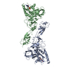
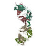
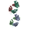
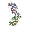
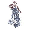
- PDB-1n0x: Crystal Structure of a Broadly Neutralizing Anti-HIV-1 Antibody i... -
+ Open data

- Basic information
| Entry | Database: PDB / ID: 1n0x | ||||||
|---|---|---|---|---|---|---|---|
| Title | Crystal Structure of a Broadly Neutralizing Anti-HIV-1 Antibody in Complex with a Peptide Mimotope | ||||||
 Components Components |
| ||||||
 Keywords Keywords | IMMUNE SYSTEM / antibody-peptide complex / peptide dimer | ||||||
| Function / homology |  Function and homology information Function and homology informationIgD immunoglobulin complex / IgA immunoglobulin complex / IgM immunoglobulin complex / IgE immunoglobulin complex / CD22 mediated BCR regulation / Fc epsilon receptor (FCERI) signaling / Classical antibody-mediated complement activation / Initial triggering of complement / IgG immunoglobulin complex / immunoglobulin mediated immune response ...IgD immunoglobulin complex / IgA immunoglobulin complex / IgM immunoglobulin complex / IgE immunoglobulin complex / CD22 mediated BCR regulation / Fc epsilon receptor (FCERI) signaling / Classical antibody-mediated complement activation / Initial triggering of complement / IgG immunoglobulin complex / immunoglobulin mediated immune response / FCGR activation / Role of LAT2/NTAL/LAB on calcium mobilization / Role of phospholipids in phagocytosis / immunoglobulin complex / Scavenging of heme from plasma / antigen binding / FCERI mediated Ca+2 mobilization / FCGR3A-mediated IL10 synthesis / Regulation of Complement cascade / Antigen activates B Cell Receptor (BCR) leading to generation of second messengers / Cell surface interactions at the vascular wall / B cell receptor signaling pathway / FCGR3A-mediated phagocytosis / FCERI mediated MAPK activation / Regulation of actin dynamics for phagocytic cup formation / Immunoregulatory interactions between a Lymphoid and a non-Lymphoid cell / FCERI mediated NF-kB activation / blood microparticle / Potential therapeutics for SARS / adaptive immune response / immune response / : / extracellular exosome / extracellular region / metal ion binding / plasma membrane Similarity search - Function | ||||||
| Biological species |  Homo sapiens (human) Homo sapiens (human)synthetic construct (others) | ||||||
| Method |  X-RAY DIFFRACTION / SYNCHROTRON / MOLECULAR REPLACEMENT / Resolution: 1.8 Å X-RAY DIFFRACTION / SYNCHROTRON / MOLECULAR REPLACEMENT / Resolution: 1.8 Å | ||||||
 Authors Authors | Saphire, E.O. / Montero, M. / Menendez, A. / Irving, M.B. / Zwick, M.B. / Parren, P.W.H.I. / Burton, D.R. / Scott, J.K. / Wilson, I.A. | ||||||
 Citation Citation | Journal: To be Published Title: Crystal Structure of a Broadly Neutralizing Anti-HIV-1 Antibody in Complex with a Peptide Mimotope Authors: Saphire, E.O. / Montero, M. / Menendez, A. / Irving, M.B. / Zwick, M.B. / Parren, P.W.H.I. / Burton, D.R. / Scott, J.K. / Wilson, I.A. | ||||||
| History |
| ||||||
| Remark 999 | Sequence The Genbank database sequence (AAA52919) is the correct match for the variable portion of ...Sequence The Genbank database sequence (AAA52919) is the correct match for the variable portion of the heavy chain except that the N-terminus has been changed from LEQSGAE to QVQLVQSGAE in the process of cloning from the recombinant Fab fragment to the IgG. The constant domains of the heavy chains H and K have the same sequence as all human IgG1 antibodies. The variable region of the light chain matches the Genbank database sequence (AAA52920) except that the N-terminus was changed in cloning to the IgG from ELTQAPG to EIVLTQSPG; the constant domains of the light chains L and M have the same sequence as all human kappa light chains. The authors maintain that the sequence of L and M, residue ALA 34 should be an ALA and not ARG (residue 33 in the sequence database). |
- Structure visualization
| Structure viewer | Molecule: MolmilJmol/JSmol |
|---|
- Downloads & links
-Download
| PDBx/mmCIF format | 1n0x.cif.gz | 212.4 KB | Display | PDBx/mmCIF format |
|---|---|---|---|---|
| PDB format | pdb1n0x.ent.gz | 166.2 KB | Display | PDB format |
| PDBx/mmJSON format | 1n0x.json.gz | Tree view | PDBx/mmJSON format | |
| Others |  Other downloads Other downloads |
-Validation report
| Arichive directory | https://data.pdbj.org/pub/pdb/validation_reports/n0/1n0xftp://data.pdbj.org/pub/pdb/validation_reports/n0/1n0x | HTTPS FTP |
|---|
-Related structure data
| Related structure data | 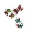 1hzhS S: Starting model for refinement |
|---|---|
| Similar structure data |


-Links
 PDBj
PDBj


- Assembly
| Deposited unit | 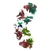
| ||||||||
|---|---|---|---|---|---|---|---|---|---|
| 1 |
| ||||||||
| Unit cell |
| ||||||||
| Details | Fab #1 is comprised of chains L and H. Fab #2 is comprised of chains M and K. / The peptide chains P and R form a dimer. Peptide chain P binds Fab #1 (LH) Peptide chain R binds Fab #2 (MK) |
-Components
-Protein/peptide , 1 types, 2 molecules PR
| #3: Protein/peptide | Mass: 2546.900 Da / Num. of mol.: 2 / Source method: obtained synthetically Details: selected from phage display peptide library, then chemically synthesized. Source: (synth.) synthetic construct (others) |
|---|
-Antibody , 2 types, 4 molecules LMHK
| #1: Antibody | Mass: 23707.354 Da / Num. of mol.: 2 / Fragment: UNP residues 132-239 Source method: isolated from a genetically manipulated source Source: (gene. exp.) Homo sapiens (human) / Cell (production host): ovary cells / Production host:   Cricetulus griseus (Chinese hamster) / Strain (production host): CHO K1 / References: UniProt: Q8TCD0, UniProt: P01834*PLUS Cricetulus griseus (Chinese hamster) / Strain (production host): CHO K1 / References: UniProt: Q8TCD0, UniProt: P01834*PLUS#2: Antibody | Mass: 24938.898 Da / Num. of mol.: 2 / Fragment: UNP residues 117-222 Source method: isolated from a genetically manipulated source Source: (gene. exp.) Homo sapiens (human) / Cell (production host): ovary cells / Production host: Cricetulus griseus (Chinese hamster) / Strain (production host): CHO K1 / References: UniProt: P0DOX5 |
|---|
-Non-polymers , 5 types, 739 molecules 


| #4: Chemical | ChemComp-GOL /  Mass: 92.094 Da / Num. of mol.: 8 / Source method: obtained synthetically / Formula: C3H8O3 Mass: 92.094 Da / Num. of mol.: 8 / Source method: obtained synthetically / Formula: C3H8O3#5: Chemical | ChemComp-SO4 /  Mass: 96.063 Da / Num. of mol.: 4 / Source method: obtained synthetically / Formula: SO4 Mass: 96.063 Da / Num. of mol.: 4 / Source method: obtained synthetically / Formula: SO4#6: Chemical | ChemComp-CXS / |  Mass: 221.317 Da / Num. of mol.: 1 / Source method: obtained synthetically / Formula: C9H19NO3S / Comment: pH buffer*YM Mass: 221.317 Da / Num. of mol.: 1 / Source method: obtained synthetically / Formula: C9H19NO3S / Comment: pH buffer*YM#7: Chemical | ChemComp-K / |  Mass: 39.098 Da / Num. of mol.: 1 / Source method: obtained synthetically / Formula: K Mass: 39.098 Da / Num. of mol.: 1 / Source method: obtained synthetically / Formula: K#8: Water | ChemComp-HOH / | Mass: 18.015 Da / Num. of mol.: 725 / Source method: isolated from a natural source / Formula: H2O |
|---|
-Details
| Has protein modification | Y |
|---|
-Experimental details
-Experiment
| Experiment | Method: X-RAY DIFFRACTION / Number of used crystals: 1 |
|---|
- Sample preparation
| Crystal | Density Matthews: 2.54 Å3/Da / Density % sol: 51.61 % |
|---|---|
| Crystal grow | Temperature: 295.5 K / Method: vapor diffusion, sitting drop / pH: 10.5 Details: ammonium sulfate, lithium sulfate, CAPS buffer, pH 10.5, VAPOR DIFFUSION, SITTING DROP, temperature 295.5K |
-Data collection
| Diffraction | Mean temperature: 171 K |
|---|---|
| Diffraction source | Source: SYNCHROTRON / Site: SSRL  / Beamline: BL11-1 / Wavelength: 0.965 Å / Beamline: BL11-1 / Wavelength: 0.965 Å |
| Detector | Type: ADSC QUANTUM 315 / Detector: CCD / Date: May 22, 2000 / Details: flat mirror |
| Radiation | Monochromator: Si 111 CHANNEL / Protocol: SINGLE WAVELENGTH / Monochromatic (M) / Laue (L): M / Scattering type: x-ray |
| Radiation wavelength | Wavelength: 0.965 Å / Relative weight: 1 |
| Reflection | Resolution: 1.78→33.45 Å / Num. all: 94383 / Num. obs: 84895 / % possible obs: 92 % / Observed criterion σ(F): 2 / Observed criterion σ(I): 2 / Redundancy: 2.3 % / Biso Wilson estimate: 20.1 Å2 / Rsym value: 0.06 / Net I/σ(I): 17.6 |
| Reflection shell | Resolution: 1.78→1.81 Å / Mean I/σ(I) obs: 1.5 / Num. unique all: 4468 / Rsym value: 0.385 / % possible all: 87.1 |
- Processing
| Software |
| |||||||||||||||||||||||||
|---|---|---|---|---|---|---|---|---|---|---|---|---|---|---|---|---|---|---|---|---|---|---|---|---|---|---|
| Refinement | Method to determine structure: MOLECULAR REPLACEMENT Starting model: Fab domains from uncomplexed IgG1 b12 structure (1HZH) Resolution: 1.8→45 Å / Cross valid method: THROUGHOUT / σ(F): 0 / Stereochemistry target values: Engh & Huber Details: 10% test set (9419) reflections selected for initial rounds of refinement. For final round of refinement, a 2% test set was randomly selected from reflections contained in the original 10% test set.
| |||||||||||||||||||||||||
| Displacement parameters |
| |||||||||||||||||||||||||
| Refinement step | Cycle: LAST / Resolution: 1.8→45 Å
|
