 Movie
Movie Controller

[English] 日本語
 Yorodumi
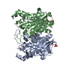
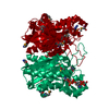
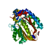
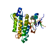
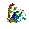
- PDB-1mzs: CRYSTAL STRUCTURE OF BETA-KETOACYL-ACP SYNTHASE III WITH BOUND di... -
+ Open data

- Basic information
| Entry | Database: PDB / ID: 1mzs | ||||||
|---|---|---|---|---|---|---|---|
| Title | CRYSTAL STRUCTURE OF BETA-KETOACYL-ACP SYNTHASE III WITH BOUND dichlorobenzyloxy-indole-carboxylic acid inhibitor | ||||||
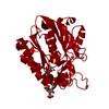
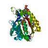
 Components Components | 3-oxoacyl-[acyl-carrier-protein] synthase III | ||||||
 Keywords Keywords | TRANSFERASE / FABH | ||||||
| Function / homology |  Function and homology information Function and homology informationbeta-ketoacyl-[acyl-carrier-protein] synthase III / beta-ketoacyl-acyl-carrier-protein synthase III activity / 3-oxoacyl-[acyl-carrier-protein] synthase activity / fatty acid metabolic process / fatty acid biosynthetic process / cytosol Similarity search - Function | ||||||
| Biological species |  | ||||||
| Method |  X-RAY DIFFRACTION / SYNCHROTRON / MOLECULAR REPLACEMENT / Resolution: 2.1 Å X-RAY DIFFRACTION / SYNCHROTRON / MOLECULAR REPLACEMENT / Resolution: 2.1 Å | ||||||
 Authors Authors | Daines, R.A. / Pendrak, I. / Sham, K. / Van Aller, G.S. / Konstantinidis, A.K. / Lonsdale, J.T. / Janson, C.A. / Qui, X. / Brandt, M. / Silverman, C. / Head, M.S. | ||||||
 Citation Citation | Journal: J.Med.Chem. / Year: 2003 Title: First X-ray cocrystal structure of a bacterial FabH condensing enzyme and a small molecule inhibitor achieved using rational design and homology modeling Authors: Daines, R.A. / Pendrak, I. / Sham, K. / Van Aller, G.S. / Konstantinidis, A.K. / Lonsdale, J.T. / Janson, C.A. / Qiu, X. / Brandt, M. / Khandekar, S.S. / Silverman, C. / Head, M.S. | ||||||
| History |
|
- Structure visualization
| Structure viewer | Molecule: MolmilJmol/JSmol |
|---|
- Downloads & links
-Download
| PDBx/mmCIF format | 1mzs.cif.gz | 75.8 KB | Display | PDBx/mmCIF format |
|---|---|---|---|---|
| PDB format | pdb1mzs.ent.gz | 56 KB | Display | PDB format |
| PDBx/mmJSON format | 1mzs.json.gz | Tree view | PDBx/mmJSON format | |
| Others |  Other downloads Other downloads |
-Validation report
| Arichive directory | https://data.pdbj.org/pub/pdb/validation_reports/mz/1mzsftp://data.pdbj.org/pub/pdb/validation_reports/mz/1mzs | HTTPS FTP |
|---|
-Related structure data
| Related structure data |  1d9b S: Starting model for refinement |
|---|---|
| Similar structure data |


-Links
 PDBj
PDBj

- Assembly
| Deposited unit | 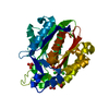
| ||||||||
|---|---|---|---|---|---|---|---|---|---|
| 1 |
| ||||||||
| 2 | 
| ||||||||
| Unit cell |
|
-Components
| #1: Protein | Mass: 33595.023 Da / Num. of mol.: 1 Source method: isolated from a genetically manipulated source Source: (gene. exp.) References: UniProt: P0A6R0, beta-ketoacyl-[acyl-carrier-protein] synthase I |
|---|---|
| #2: Chemical | ChemComp-PO4 /   Mass: 94.971 Da / Num. of mol.: 1 / Source method: obtained synthetically / Formula: PO4 Mass: 94.971 Da / Num. of mol.: 1 / Source method: obtained synthetically / Formula: PO4 |
| #3: Chemical | ChemComp-669 / 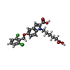  Mass: 450.312 Da / Num. of mol.: 1 / Source method: obtained synthetically / Formula: C22H21Cl2NO5 Mass: 450.312 Da / Num. of mol.: 1 / Source method: obtained synthetically / Formula: C22H21Cl2NO5 |
| #4: Water | ChemComp-HOH /  Mass: 18.015 Da / Num. of mol.: 177 / Source method: isolated from a natural source / Formula: H2O Mass: 18.015 Da / Num. of mol.: 177 / Source method: isolated from a natural source / Formula: H2O |
| Has protein modification | Y |
-Experimental details
-Experiment
| Experiment | Method: X-RAY DIFFRACTION / Number of used crystals: 1 |
|---|
- Sample preparation
| Crystal | Density Matthews: 1.96 Å3/Da / Density % sol: 37.27 % |
|---|---|
| Crystal grow | Temperature: 278 K / Method: vapor diffusion, sitting drop / pH: 7 Details: 20% PEG 8000, pH 7.0, VAPOR DIFFUSION, SITTING DROP, temperature 278K |
| Crystal grow | *PLUS Temperature: 4 ℃ / pH: 7 / Method: unknown / Details: Qiu, X., (1999) J.Biol.Chem., 274, 36465. |
| Components of the solutions | *PLUS Conc.: 14 % / Common name: PEG6000 |
-Data collection
| Diffraction | Mean temperature: 100 K |
|---|---|
| Diffraction source | Source: SYNCHROTRON / Site: APS  / Beamline: 17-ID / Wavelength: 1 Å / Beamline: 17-ID / Wavelength: 1 Å |
| Detector | Type: MARRESEARCH / Detector: CCD / Date: Mar 1, 2000 |
| Radiation | Monochromator: Si 111 / Protocol: SINGLE WAVELENGTH / Monochromatic (M) / Laue (L): M / Scattering type: x-ray |
| Radiation wavelength | Wavelength: 1 Å / Relative weight: 1 |
| Reflection | Resolution: 2.1→20 Å / Num. all: 14354 / Num. obs: 14354 / % possible obs: 85.3 % / Observed criterion σ(F): 0 / Observed criterion σ(I): 0 / Redundancy: 7.9 % / Biso Wilson estimate: 26 Å2 / Rmerge(I) obs: 0.111 / Rsym value: 0.111 / Net I/σ(I): 15 |
| Reflection shell | Resolution: 2.1→2.17 Å / Redundancy: 3 % / Rmerge(I) obs: 0.301 / Mean I/σ(I) obs: 3.15 / Num. unique all: 1572 / Rsym value: 0.301 / % possible all: 0.92 |
| Reflection | *PLUS Lowest resolution: 20 Å |
- Processing
| Software |
| ||||||||||||||||||||||||||||
|---|---|---|---|---|---|---|---|---|---|---|---|---|---|---|---|---|---|---|---|---|---|---|---|---|---|---|---|---|---|
| Refinement | Method to determine structure: MOLECULAR REPLACEMENT Starting model: pdb entry 1d9b 1d9b Resolution: 2.1→20 Å / Cross valid method: THROUGHOUT / σ(F): 0 / σ(I): 0 / Stereochemistry target values: Engh & Huber
| ||||||||||||||||||||||||||||
| Refinement step | Cycle: LAST / Resolution: 2.1→20 Å
| ||||||||||||||||||||||||||||
| Refine LS restraints |
| ||||||||||||||||||||||||||||
| LS refinement shell | Resolution: 2.1→2.19 Å / Total num. of bins used: 10
| ||||||||||||||||||||||||||||
| Refinement | *PLUS Lowest resolution: 20 Å / Rfactor Rfree: 0.23 | ||||||||||||||||||||||||||||
| Solvent computation | *PLUS | ||||||||||||||||||||||||||||
| Displacement parameters | *PLUS |
