 Movie
Movie Controller


+ Open data

- Basic information
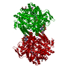
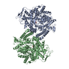
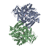
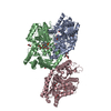
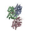
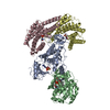
| Entry | Database: PDB / ID: 1mzo | |||||||||
|---|---|---|---|---|---|---|---|---|---|---|
| Title | Crystal structure of pyruvate formate-lyase with pyruvate | |||||||||
 Components Components | Pyruvate formate-lyase | |||||||||
 Keywords Keywords | TRANSFERASE / ENZYME-SUBSTRATE COMPLEX | |||||||||
| Function / homology |  Function and homology information Function and homology information: / formate C-acetyltransferase / formate C-acetyltransferase activity / glucose metabolic process / membrane / cytosol Similarity search - Function | |||||||||
| Biological species |  | |||||||||
| Method |  X-RAY DIFFRACTION / SYNCHROTRON / MOLECULAR REPLACEMENT / Resolution: 2.7 Å X-RAY DIFFRACTION / SYNCHROTRON / MOLECULAR REPLACEMENT / Resolution: 2.7 Å | |||||||||
 Authors Authors | Lehtio, L. / Leppanen, V.-M. / Kozarich, J.W. / Goldman, A. | |||||||||
 Citation Citation | Journal: Acta Crystallogr.,Sect.D / Year: 2002 Title: Structure of Escherichia coli pyruvate formate-lyase with pyruvate. Authors: Lehtio, L. / Leppanen, V.M. / Kozarich, J.W. / Goldman, A. | |||||||||
| History |
|
- Structure visualization
| Structure viewer | Molecule: MolmilJmol/JSmol |
|---|
- Downloads & links
-Download
| PDBx/mmCIF format | 1mzo.cif.gz | 307.3 KB | Display | PDBx/mmCIF format |
|---|---|---|---|---|
| PDB format | pdb1mzo.ent.gz | 250.6 KB | Display | PDB format |
| PDBx/mmJSON format | 1mzo.json.gz | Tree view | PDBx/mmJSON format | |
| Others |  Other downloads Other downloads |
-Validation report
| Arichive directory | https://data.pdbj.org/pub/pdb/validation_reports/mz/1mzoftp://data.pdbj.org/pub/pdb/validation_reports/mz/1mzo | HTTPS FTP |
|---|
-Related structure data
| Related structure data | 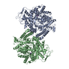 2pflS S: Starting model for refinement |
|---|---|
| Similar structure data |


-Links
 PDBj
PDBj

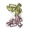
- Assembly
| Deposited unit | 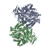
| ||||||||
|---|---|---|---|---|---|---|---|---|---|
| 1 |
| ||||||||
| Unit cell |
| ||||||||
| Components on special symmetry positions |
|
-Components
| #1: Protein | Mass: 85327.898 Da / Num. of mol.: 2 Source method: isolated from a genetically manipulated source Source: (gene. exp.) #2: Chemical | ChemComp-PGE / |   Mass: 150.173 Da / Num. of mol.: 1 / Source method: obtained synthetically / Formula: C6H14O4 Mass: 150.173 Da / Num. of mol.: 1 / Source method: obtained synthetically / Formula: C6H14O4#3: Chemical |   Mass: 88.062 Da / Num. of mol.: 2 / Source method: obtained synthetically / Formula: C3H4O3 Mass: 88.062 Da / Num. of mol.: 2 / Source method: obtained synthetically / Formula: C3H4O3#4: Water | ChemComp-HOH / |  Mass: 18.015 Da / Num. of mol.: 515 / Source method: isolated from a natural source / Formula: H2O Mass: 18.015 Da / Num. of mol.: 515 / Source method: isolated from a natural source / Formula: H2O |
|---|
-Experimental details
-Experiment
| Experiment | Method: X-RAY DIFFRACTION / Number of used crystals: 1 |
|---|
- Sample preparation
| Crystal | Density Matthews: 2.92 Å3/Da / Density % sol: 57.95 % | |||||||||||||||||||||||||||||||||||||||||||||||||||||||||||||||||||||||||||||
|---|---|---|---|---|---|---|---|---|---|---|---|---|---|---|---|---|---|---|---|---|---|---|---|---|---|---|---|---|---|---|---|---|---|---|---|---|---|---|---|---|---|---|---|---|---|---|---|---|---|---|---|---|---|---|---|---|---|---|---|---|---|---|---|---|---|---|---|---|---|---|---|---|---|---|---|---|---|---|
| Crystal grow | Temperature: 298 K / Method: microdialysis / pH: 7.3 Details: PEG1000, MOPS, ammonium chloride, na-pyruvate, DTT, magnesium chloride, EDTA, pH 7.3, MICRODIALYSIS, temperature 298K | |||||||||||||||||||||||||||||||||||||||||||||||||||||||||||||||||||||||||||||
| Crystal grow | *PLUS | |||||||||||||||||||||||||||||||||||||||||||||||||||||||||||||||||||||||||||||
| Components of the solutions | *PLUS
|
-Data collection
| Diffraction | Mean temperature: 100 K |
|---|---|
| Diffraction source | Source: SYNCHROTRON / Site: EMBL/DESY, HAMBURG  / Beamline: X11 / Wavelength: 0.9102 Å / Beamline: X11 / Wavelength: 0.9102 Å |
| Detector | Type: MARRESEARCH / Detector: CCD / Date: Jun 20, 2000 |
| Radiation | Protocol: SINGLE WAVELENGTH / Monochromatic (M) / Laue (L): M / Scattering type: x-ray |
| Radiation wavelength | Wavelength: 0.9102 Å / Relative weight: 1 |
| Reflection | Resolution: 2.7→20 Å / Num. all: 56141 / Num. obs: 55923 / % possible obs: 98.8 % / Observed criterion σ(F): -1 |
| Reflection shell | Resolution: 2.7→2.8 Å / % possible all: 94.9 |
| Reflection | *PLUS Lowest resolution: 20 Å / Num. measured all: 318766 / Rmerge(I) obs: 0.111 |
| Reflection shell | *PLUS % possible obs: 94.9 % / Rmerge(I) obs: 0.34 / Mean I/σ(I) obs: 1.5 |
- Processing
| Software |
| ||||||||||||||||||||||||
|---|---|---|---|---|---|---|---|---|---|---|---|---|---|---|---|---|---|---|---|---|---|---|---|---|---|
| Refinement | Method to determine structure: MOLECULAR REPLACEMENT Starting model: PDB ENTRY 2PFL Resolution: 2.7→20 Å / Cross valid method: THROUGHOUT / σ(F): 0 / Stereochemistry target values: Engh & Huber Details: 9 OF THE POLYETHYLENE GLYCOL ATOMS WERE INCLUDED IN REFINEMENT AND ATOM O1 ADDED AFTERWARDS ON SYMMETRY AXIS WITH 0.5 OCCUPANCY
| ||||||||||||||||||||||||
| Refinement step | Cycle: LAST / Resolution: 2.7→20 Å
| ||||||||||||||||||||||||
| Refinement | *PLUS Lowest resolution: 20 Å / % reflection Rfree: 5 % | ||||||||||||||||||||||||
| Solvent computation | *PLUS | ||||||||||||||||||||||||
| Displacement parameters | *PLUS | ||||||||||||||||||||||||
| Refine LS restraints | *PLUS
|
