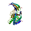
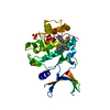
 Movie
Movie Controller


+ Open data

- Basic information
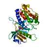
| Entry | Database: PDB / ID: 1mwj | ||||||
|---|---|---|---|---|---|---|---|
| Title | Crystal Structure of a MUG-DNA pseudo substrate complex | ||||||
 Components Components |
| ||||||
 Keywords Keywords | HYDROLASE/DNA / Rossmann Fold / non-hydrolysable DNA-complex / Uracil recognition. / HYDROLASE-DNA COMPLEX | ||||||
| Function / homology |  Function and homology information Function and homology informationdouble-stranded uracil-DNA glycosylase / pyrimidine-specific mismatch base pair DNA N-glycosylase activity / base-excision repair, AP site formation / uracil DNA N-glycosylase activity / DNA binding / cytoplasm Similarity search - Function | ||||||
| Biological species |  | ||||||
| Method |  X-RAY DIFFRACTION / SYNCHROTRON / MOLECULAR REPLACEMENT / Resolution: 2.85 Å X-RAY DIFFRACTION / SYNCHROTRON / MOLECULAR REPLACEMENT / Resolution: 2.85 Å | ||||||
 Authors Authors | Barrett, T.E. / Scharer, O. / Savva, R. / Brown, T. / Jiricny, J. / Verdine, G.L. / Pearl, L.H. | ||||||
 Citation Citation | Journal: Embo J. / Year: 1999 Title: Crystal Structure of a thwarted mismatch glycosylase DNA repair complex Authors: Barrett, T.E. / Scharer, O. / Savva, R. / Brown, T. / Jiricny, J. / Verdine, G.L. / Pearl, L.H. | ||||||
| History |
| ||||||
| Remark 300 | BIOMOLECULE THIS ENTRY CONTAINS THE CRYSTALLOGRAPHIC ASYMMETRIC UNIT WHICH CONSISTS OF 2 CHAIN(S). ...BIOMOLECULE THIS ENTRY CONTAINS THE CRYSTALLOGRAPHIC ASYMMETRIC UNIT WHICH CONSISTS OF 2 CHAIN(S). The second strand of the DNA molecule is generated by applying symmetry operator 7_555 : y, x, -z, to DU and bases 8-12, and applying symmetry operator 7_554 : y, x, -1-z, to bases 1-6. |
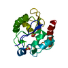
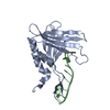
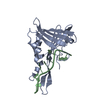
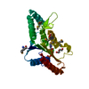
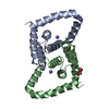
- Structure visualization
| Structure viewer | Molecule: MolmilJmol/JSmol |
|---|
- Downloads & links
-Download
| PDBx/mmCIF format | 1mwj.cif.gz | 55.5 KB | Display | PDBx/mmCIF format |
|---|---|---|---|---|
| PDB format | pdb1mwj.ent.gz | 37.1 KB | Display | PDB format |
| PDBx/mmJSON format | 1mwj.json.gz | Tree view | PDBx/mmJSON format | |
| Others |  Other downloads Other downloads |
-Validation report
| Arichive directory | https://data.pdbj.org/pub/pdb/validation_reports/mw/1mwjftp://data.pdbj.org/pub/pdb/validation_reports/mw/1mwj | HTTPS FTP |
|---|
-Related structure data
| Related structure data | |
|---|---|
| Similar structure data |


-Links
 PDBj
PDBj


- Assembly
| Deposited unit | 
| ||||||||
|---|---|---|---|---|---|---|---|---|---|
| 1 | 
| ||||||||
| Unit cell |
|
-Components
| #1: DNA chain | Mass: 3665.365 Da / Num. of mol.: 1 / Source method: obtained synthetically |
|---|---|
| #2: Protein | Mass: 18696.352 Da / Num. of mol.: 1 Source method: isolated from a genetically manipulated source Source: (gene. exp.) References: UniProt: P0A9H1, Hydrolases; Glycosylases; Hydrolysing N-glycosyl compounds |
| #3: Water | ChemComp-HOH /  Mass: 18.015 Da / Num. of mol.: 58 / Source method: isolated from a natural source / Formula: H2O Mass: 18.015 Da / Num. of mol.: 58 / Source method: isolated from a natural source / Formula: H2O |
| Has protein modification | N |
-Experimental details
-Experiment
| Experiment | Method: X-RAY DIFFRACTION / Number of used crystals: 1 |
|---|
- Sample preparation
| Crystal | Density Matthews: 2.6 Å3/Da / Density % sol: 52.78 % | |||||||||||||||||||||||||||||||||||
|---|---|---|---|---|---|---|---|---|---|---|---|---|---|---|---|---|---|---|---|---|---|---|---|---|---|---|---|---|---|---|---|---|---|---|---|---|
| Crystal grow | Temperature: 289 K / Method: microbatch / pH: 4.6 Details: PEG4000, Sodium Acetate, pH 4.6, Microbatch, temperature 289K | |||||||||||||||||||||||||||||||||||
| Components of the solutions |
| |||||||||||||||||||||||||||||||||||
| Crystal grow | *PLUS Method: microdialysis / Details: Barrett, T.E., (1998) Cell., 92, 117. | |||||||||||||||||||||||||||||||||||
| Components of the solutions | *PLUS
|
-Data collection
| Diffraction | Mean temperature: 100 K |
|---|---|
| Diffraction source | Source: SYNCHROTRON / Site: SRS  / Beamline: PX7.2 / Wavelength: 1.488 Å / Beamline: PX7.2 / Wavelength: 1.488 Å |
| Detector | Type: MARRESEARCH / Detector: IMAGE PLATE / Date: Feb 15, 1998 |
| Radiation | Monochromator: Si 111 CHANNEL / Protocol: SINGLE WAVELENGTH / Monochromatic (M) / Laue (L): M / Scattering type: x-ray |
| Radiation wavelength | Wavelength: 1.488 Å / Relative weight: 1 |
| Reflection | Resolution: 2.85→20 Å / Num. all: 5478 / Num. obs: 5478 / % possible obs: 94.5 % / Observed criterion σ(F): 0 / Observed criterion σ(I): 2 / Redundancy: 2.3 % / Biso Wilson estimate: 38.2 Å2 / Rmerge(I) obs: 0.087 / Rsym value: 0.087 / Net I/σ(I): 9.7 |
| Reflection shell | Resolution: 2.85→2.95 Å / Redundancy: 1.8 % / Rmerge(I) obs: 0.263 / Mean I/σ(I) obs: 2 / Num. unique all: 472 / Rsym value: 0.263 / % possible all: 85.7 |
| Reflection | *PLUS Num. obs: 5479 / Rmerge(I) obs: 0.087 |
| Reflection shell | *PLUS % possible obs: 96.7 % / Rmerge(I) obs: 0.278 / Mean I/σ(I) obs: 2.9 |
- Processing
| Software |
| ||||||||||||||||||||||||||||||||||||
|---|---|---|---|---|---|---|---|---|---|---|---|---|---|---|---|---|---|---|---|---|---|---|---|---|---|---|---|---|---|---|---|---|---|---|---|---|---|
| Refinement | Method to determine structure: MOLECULAR REPLACEMENT Starting model: Native MuG Resolution: 2.85→14.97 Å / Rfactor Rfree error: 0.017 / Data cutoff high absF: 1104323.12 / Data cutoff low absF: 0 / Isotropic thermal model: RESTRAINED / Cross valid method: THROUGHOUT / σ(F): 0 / Stereochemistry target values: Engh & Huber Details: NO ELECTRON DENSITY IS OBSERVED FOR RESDUES 166 TO 168 RESIDUES A1, A109, A150 AND A151 WERE REFINED AS ALANINE
| ||||||||||||||||||||||||||||||||||||
| Displacement parameters | Biso mean: 22.3 Å2 | ||||||||||||||||||||||||||||||||||||
| Refine analyze | Luzzati coordinate error obs: 0.35 Å / Luzzati d res low obs: 5 Å / Luzzati sigma a obs: 0.38 Å | ||||||||||||||||||||||||||||||||||||
| Refinement step | Cycle: LAST / Resolution: 2.85→14.97 Å
| ||||||||||||||||||||||||||||||||||||
| Refine LS restraints |
| ||||||||||||||||||||||||||||||||||||
| LS refinement shell | Resolution: 2.85→2.93 Å / Rfactor Rfree error: 0.018 / Total num. of bins used: 8
| ||||||||||||||||||||||||||||||||||||
| Xplor file |
| ||||||||||||||||||||||||||||||||||||
| Software | *PLUS Name: XPLOR / Version: 3.851 / Classification: refinement | ||||||||||||||||||||||||||||||||||||
| Refinement | *PLUS Lowest resolution: 20 Å / Rfactor obs: 0.23 / Rfactor Rfree: 0.25 / Rfactor Rwork: 0.19 | ||||||||||||||||||||||||||||||||||||
| Solvent computation | *PLUS | ||||||||||||||||||||||||||||||||||||
| Displacement parameters | *PLUS | ||||||||||||||||||||||||||||||||||||
| Refine LS restraints | *PLUS
| ||||||||||||||||||||||||||||||||||||
| LS refinement shell | *PLUS Rfactor Rfree: 0.367 / Rfactor Rwork: 0.295 |
