 Movie
Movie Controller

[English] 日本語
 Yorodumi
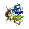
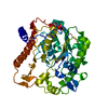
- PDB-1mqe: Structure of the MT-ADPRase in complex with gadolidium and ADP-ri... -
+ Open data

- Basic information
| Entry | Database: PDB / ID: 1mqe | ||||||
|---|---|---|---|---|---|---|---|
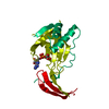
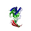
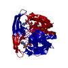
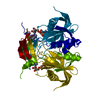
| Title | Structure of the MT-ADPRase in complex with gadolidium and ADP-ribose, a Nudix enzyme | ||||||
 Components Components | ADPR pyrophosphatase | ||||||
 Keywords Keywords | HYDROLASE / Nudix hydrolase / Rv1700 / ADPR / Mycobacterium Tuberculosis | ||||||
| Function / homology |  Function and homology information Function and homology informationnucleoside phosphate metabolic process / ribose phosphate metabolic process / manganese ion binding / hydrolase activity / cytosol Similarity search - Function | ||||||
| Biological species |   Mycobacterium tuberculosis (bacteria) Mycobacterium tuberculosis (bacteria) | ||||||
| Method |  X-RAY DIFFRACTION / MIR / Resolution: 2 Å X-RAY DIFFRACTION / MIR / Resolution: 2 Å | ||||||
 Authors Authors | Kang, L.-W. / Gabelli, S.B. / Bianchet, M.A. / Cunningham, J.E. / O'Handley, S.F. / Amzel, L.M. | ||||||
 Citation Citation | Journal: Structure / Year: 2003 Title: Structure and mechanism of MT-ADPRase, a Nudix hydrolase from Mycobacterium tuberculosis Authors: Kang, L.-W. / Gabelli, S.B. / Cunningham, J.E. / O'Handley, S.F. / Amzel, L.M. | ||||||
| History |
|
- Structure visualization
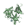
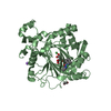
| Structure viewer | Molecule: MolmilJmol/JSmol |
|---|
- Downloads & links
-Download
| PDBx/mmCIF format | 1mqe.cif.gz | 52.3 KB | Display | PDBx/mmCIF format |
|---|---|---|---|---|
| PDB format | pdb1mqe.ent.gz | 37.3 KB | Display | PDB format |
| PDBx/mmJSON format | 1mqe.json.gz | Tree view | PDBx/mmJSON format | |
| Others |  Other downloads Other downloads |
-Validation report
| Arichive directory | https://data.pdbj.org/pub/pdb/validation_reports/mq/1mqeftp://data.pdbj.org/pub/pdb/validation_reports/mq/1mqe | HTTPS FTP |
|---|
-Related structure data


-Links
 PDBj
PDBj


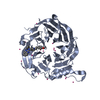
- Assembly
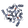
| Deposited unit | 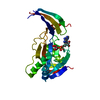
| ||||||||
|---|---|---|---|---|---|---|---|---|---|
| 1 | 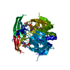
| ||||||||
| Unit cell |
| ||||||||
| Details | The second part of the biological assembly is generated by the two fold axis |
-Components
| #1: Protein | Mass: 22920.846 Da / Num. of mol.: 1 Source method: isolated from a genetically manipulated source Source: (gene. exp.) Mycobacterium tuberculosis (bacteria) / Plasmid: pET11b / Production host: |
|---|---|
| #2: Chemical | ChemComp-APR /   Mass: 559.316 Da / Num. of mol.: 1 / Source method: obtained synthetically / Formula: C15H23N5O14P2 Mass: 559.316 Da / Num. of mol.: 1 / Source method: obtained synthetically / Formula: C15H23N5O14P2 |
| #3: Chemical | ChemComp-GD3 /   Mass: 157.250 Da / Num. of mol.: 1 / Source method: obtained synthetically / Formula: Gd Mass: 157.250 Da / Num. of mol.: 1 / Source method: obtained synthetically / Formula: Gd |
| #4: Water | ChemComp-HOH /  Mass: 18.015 Da / Num. of mol.: 86 / Source method: isolated from a natural source / Formula: H2O Mass: 18.015 Da / Num. of mol.: 86 / Source method: isolated from a natural source / Formula: H2O |
-Experimental details
-Experiment
| Experiment | Method: X-RAY DIFFRACTION / Number of used crystals: 1 |
|---|
- Sample preparation
| Crystal | Density Matthews: 2.35 Å3/Da / Density % sol: 47.72 % | ||||||||||||||||||||||||||||||||||||
|---|---|---|---|---|---|---|---|---|---|---|---|---|---|---|---|---|---|---|---|---|---|---|---|---|---|---|---|---|---|---|---|---|---|---|---|---|---|
| Crystal grow | Temperature: 298 K / Method: vapor diffusion, hanging drop / pH: 7.5 Details: Sodium formate, Tris HCl, pH 7.5, VAPOR DIFFUSION, HANGING DROP, temperature 298K | ||||||||||||||||||||||||||||||||||||
| Crystal grow | *PLUS Method: vapor diffusion, hanging drop | ||||||||||||||||||||||||||||||||||||
| Components of the solutions | *PLUS
|
-Data collection
| Diffraction | Mean temperature: 93 K |
|---|---|
| Diffraction source | Source: ROTATING ANODE / Type: RIGAKU RU200 / Wavelength: 1.54 Å |
| Detector | Type: RIGAKU RAXIS IV / Detector: IMAGE PLATE / Date: Nov 20, 2001 / Details: mirrors |
| Radiation | Monochromator: Ni filter / Protocol: SINGLE WAVELENGTH / Monochromatic (M) / Laue (L): M / Scattering type: x-ray |
| Radiation wavelength | Wavelength: 1.54 Å / Relative weight: 1 |
| Reflection | Resolution: 2→30 Å / Num. all: 15835 / Num. obs: 15604 / % possible obs: 98.6 % / Observed criterion σ(F): 0 / Observed criterion σ(I): 0 / Rsym value: 0.075 / Net I/σ(I): 21.3 |
| Reflection shell | Resolution: 2→2.07 Å / Rsym value: 0.336 / % possible all: 97.5 |
| Reflection | *PLUS Num. measured all: 127268 / Rmerge(I) obs: 0.075 |
| Reflection shell | *PLUS % possible obs: 97.5 % / Mean I/σ(I) obs: 5.7 |
- Processing
| Software |
| ||||||||||||||||||||
|---|---|---|---|---|---|---|---|---|---|---|---|---|---|---|---|---|---|---|---|---|---|
| Refinement | Method to determine structure: MIR / Resolution: 2→30 Å / σ(F): 0 / Stereochemistry target values: Engh & Huber
| ||||||||||||||||||||
| Refinement step | Cycle: LAST / Resolution: 2→30 Å
| ||||||||||||||||||||
| Refine LS restraints |
| ||||||||||||||||||||
| Refinement | *PLUS | ||||||||||||||||||||
| Solvent computation | *PLUS | ||||||||||||||||||||
| Displacement parameters | *PLUS | ||||||||||||||||||||
| Refine LS restraints | *PLUS
|
