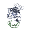
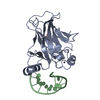
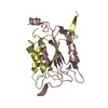
double-stranded uracil-DNA glycosylase / pyrimidine-specific mismatch base pair DNA N-glycosylase activity / base-excision repair, AP site formation / uracil DNA N-glycosylase activity / DNA binding / cytoplasm Similarity search - Function
G/U mismatch-specific DNA glycosylase MUG, bacterial / Uracil DNA glycosylase family 2 / Uracil-DNA Glycosylase, subunit E / Uracil-DNA glycosylase-like domain / Uracil-DNA glycosylase-like / Uracil DNA glycosylase superfamily / Uracil-DNA glycosylase-like domain superfamily / 3-Layer(aba) Sandwich / Alpha Beta Similarity search - Domain/homology
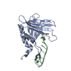
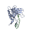
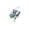
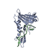
BIOMOLECULE THIS ENTRY CONTAINS THE CRYSTALLOGRAPHIC ASYMMETRIC UNIT WHICH CONSISTS OF 2 CHAIN(S). ...BIOMOLECULE THIS ENTRY CONTAINS THE CRYSTALLOGRAPHIC ASYMMETRIC UNIT WHICH CONSISTS OF 2 CHAIN(S). The second strand of the DNA molecule is generated by applying symmetry operator 7_556 : y, x, 1-z, to D1P and bases 8-12, and applying symmetry operator 7_557 : y, x, 2-z, to bases 1-6.
One asymmetric unit consists of a single protein molecule and 1 strand of DNA. The second strand is generated by x,y,-z (translation 0,0,1) applied to APR,THY,CYT,GUA, CYT,GUA and x,y,-z (translation 0,0,2) applied to the remaining nucleic acids.
-
Components
#1: DNA chain
5'-D(*CP*GP*CP*GP*AP*GP*(AAB)P*TP*CP*GP*CP*G)-3'
Mass: 3571.293 Da / Num. of mol.: 1 / Source method: obtained synthetically
Resolution: 2.35→2.55 Å / Redundancy: 4.5 % / Rmerge(I) obs: 0.2 / Rsym value: 0.2 / % possible all: 95.2
-
Processing
Software
Name
Version
Classification
DENZO
datareduction
SCALEPACK
datascaling
AMoRE
phasing
X-PLOR
3.851
refinement
Refinement
Method to determine structure: MOLECULAR REPLACEMENT Starting model: Native MUG Resolution: 2.35→14.93 Å / Rfactor Rfree error: 0.015 / Data cutoff high absF: 1604690.93 / Data cutoff low absF: 0 / Isotropic thermal model: RESTRAINED / Cross valid method: THROUGHOUT / σ(F): 0 / Stereochemistry target values: Engh & Huber Details: NO ELECTRON DENSITY IS OBSERVED FOR RESDUES 166 TO 168. RESIDUES A1, A109, A150 AND A151 WERE REFINED AS ALANINE. D1P CORRESPONDS TO AN ABASIC SITE
Rfactor
Num. reflection
% reflection
Selection details
Rfree
0.28
477
4.8 %
RANDOM
Rwork
0.21
-
-
-
all
0.2482
10144
-
-
obs
0.2482
9878
99.2 %
-
Displacement parameters
Biso mean: 23.4 Å2
Baniso -1
Baniso -2
Baniso -3
1-
0 Å2
0 Å2
0 Å2
2-
-
0 Å2
0 Å2
3-
-
-
0 Å2
Refine analyze
Luzzati coordinate error obs: 0.3 Å / Luzzati d res low obs: 5 Å / Luzzati sigma a obs: 0.37 Å
Refinement step
Cycle: LAST / Resolution: 2.35→14.93 Å
Protein
Nucleic acid
Ligand
Solvent
Total
Num. atoms
1271
236
0
122
1629
Refine LS restraints
Refine-ID
Type
Dev ideal
Dev ideal target
X-RAY DIFFRACTION
x_bond_d
0.007
X-RAY DIFFRACTION
x_angle_deg
1.1
X-RAY DIFFRACTION
x_dihedral_angle_d
26.5
X-RAY DIFFRACTION
x_improper_angle_d
1.19
X-RAY DIFFRACTION
x_mcbond_it
1.62
1.5
X-RAY DIFFRACTION
x_mcangle_it
2.53
2
X-RAY DIFFRACTION
x_scbond_it
3.5
2
X-RAY DIFFRACTION
x_scangle_it
4.8
2.5
LS refinement shell
Resolution: 2.35→2.5 Å / Rfactor Rfree error: 0.042 / Total num. of bins used: 8
Rfactor
Num. reflection
% reflection
Rfree
0.29
64
5.3 %
Rwork
0.28
1102
-
obs
-
-
95.1 %
Xplor file
Refine-ID
Serial no
Param file
Topol file
X-RAY DIFFRACTION
1
PROTEIN_REP.PARAM
TOPHCSDX.PRO
X-RAY DIFFRACTION
2
WATER.PAR
WATER.TOP
X-RAY DIFFRACTION
3
DNA2.PAR
DNA2.TOP
+
About Yorodumi
-
News
-
Feb 9, 2022. New format data for meta-information of EMDB entries
New format data for meta-information of EMDB entries
Version 3 of the EMDB header file is now the official format.
The previous official version 1.9 will be removed from the archive.
In the structure databanks used in Yorodumi, some data are registered as the other names, "COVID-19 virus" and "2019-nCoV". Here are the details of the virus and the list of structure data.
Jan 31, 2019. EMDB accession codes are about to change! (news from PDBe EMDB page)
EMDB accession codes are about to change! (news from PDBe EMDB page)
The allocation of 4 digits for EMDB accession codes will soon come to an end. Whilst these codes will remain in use, new EMDB accession codes will include an additional digit and will expand incrementally as the available range of codes is exhausted. The current 4-digit format prefixed with “EMD-” (i.e. EMD-XXXX) will advance to a 5-digit format (i.e. EMD-XXXXX), and so on. It is currently estimated that the 4-digit codes will be depleted around Spring 2019, at which point the 5-digit format will come into force.
The EM Navigator/Yorodumi systems omit the EMD- prefix.
Related info.:Q: What is EMD? / ID/Accession-code notation in Yorodumi/EM Navigator
Yorodumi is a browser for structure data from EMDB, PDB, SASBDB, etc.
This page is also the successor to EM Navigator detail page, and also detail information page/front-end page for Omokage search.
The word "yorodu" (or yorozu) is an old Japanese word meaning "ten thousand". "mi" (miru) is to see.
Related info.:EMDB / PDB / SASBDB / Comparison of 3 databanks / Yorodumi Search / Aug 31, 2016. New EM Navigator & Yorodumi / Yorodumi Papers / Jmol/JSmol / Function and homology information / Changes in new EM Navigator and Yorodumi
 Movie
Movie Controller


 Open data
 Basic information
 Components
 Keywords
 Function and homology information
 X-RAY DIFFRACTION /
  Authors
 Citation
 Structure visualization
 Downloads & links
 Other downloads
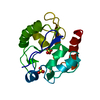


 PDBj
PDBj


 Assembly
 Mass: 18.015 Da / Num. of mol.: 122 / Source method: isolated from a natural source / Formula: H2O
 Sample preparation
 / Beamline: PX7.2 / Wavelength: 1.488 Å
 Processing