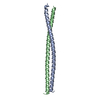
 Movie
Movie Controller

[English] 日本語
 Yorodumi
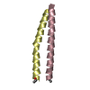
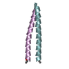
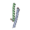
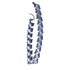
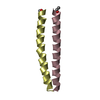
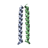
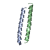
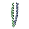
- PDB-1mv4: TM9A251-284: A Peptide Model of the C-Terminus of a Rat Striated ... -
+ Open data

- Basic information
| Entry | Database: PDB / ID: 1mv4 | ||||||
|---|---|---|---|---|---|---|---|
| Title | TM9A251-284: A Peptide Model of the C-Terminus of a Rat Striated Alpha Tropomyosin | ||||||
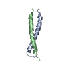
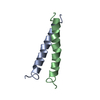
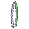
 Components Components | Tropomyosin 1 alpha chain | ||||||
 Keywords Keywords | DE NOVO PROTEIN / TROPOMYOSIN / EXON 9A / ACTIN-BINDING / TROPONIN BINDING / MUSCLE / ALPHA-HELIX / COILED-COIL / DIMER / PEPTIDE-MODEL / TWO-CHAINED / DISULFIDE CROSS-LINKED | ||||||
| Function / homology |  Function and homology information Function and homology informationStriated Muscle Contraction / Smooth Muscle Contraction / positive regulation of heart rate by epinephrine / bleb / ruffle organization / actin filament capping / muscle filament sliding / sarcomere organization / ventricular cardiac muscle tissue morphogenesis / negative regulation of vascular associated smooth muscle cell migration ...Striated Muscle Contraction / Smooth Muscle Contraction / positive regulation of heart rate by epinephrine / bleb / ruffle organization / actin filament capping / muscle filament sliding / sarcomere organization / ventricular cardiac muscle tissue morphogenesis / negative regulation of vascular associated smooth muscle cell migration / myofibril / negative regulation of vascular associated smooth muscle cell proliferation / cytoskeletal protein binding / cardiac muscle contraction / positive regulation of stress fiber assembly / stress fiber / muscle contraction / positive regulation of cell adhesion / negative regulation of cell migration / actin filament organization / actin filament / wound healing / cellular response to reactive oxygen species / ruffle membrane / disordered domain specific binding / actin filament binding / regulation of cell shape / actin cytoskeleton / in utero embryonic development / actin binding / protein heterodimerization activity / protein homodimerization activity / protein-containing complex / identical protein binding / cytosol / cytoplasm Similarity search - Function | ||||||
| Biological species |  | ||||||
| Method | SOLUTION NMR / 1648 CONFORMATIONALLY- RESTRICTING NOE CONSTRAINTS (OUT OF A TOTAL OF 2036 NOE DERIVED DISTANCES) 52 LOOSE DIHEDRAL ANGLE CONSTRAINTS, 48 BACKBONE INTRA- HELICAL HYDROGEN BOND CONSTRAINTS WERE UTILILIZED. | ||||||
 Authors Authors | Greenfield, N.J. / Swapna, G.V.T. / Huang, Y. / Palm, T. / Graboski, S. / Montelione, G.T. / Hitchcock-Degregori, S.E. | ||||||
 Citation Citation | Journal: Biochemistry / Year: 2003 Title: The Structure of the Carboxyl Terminus of Striated alpha-Tropomyosin in Solution Reveals an Unusual Parallel Arrangement of Interacting alpha-Helices Authors: Greenfield, N.J. / Swapna, G.V.T. / Huang, Y. / Palm, T. / Graboski, S. / Montelione, G.T. / Hitchcock-DeGregori, S.E. | ||||||
| History |
| ||||||
| Remark 999 | SEQUENCE THIS SEQUENCE HAS A SYNTHETIC GCG AT N-TERMINUS AND CONTAINS THE MUTATION N279K. THE ...SEQUENCE THIS SEQUENCE HAS A SYNTHETIC GCG AT N-TERMINUS AND CONTAINS THE MUTATION N279K. THE SEQUENCE DATABASE MATCH, HOWEVER, erroneously HAS A K AT THIS POSITION as well. The sequence in the database should have an ASN at position 279, which has been engineered to LYS in this entry. |
- Structure visualization
| Structure viewer | Molecule: MolmilJmol/JSmol |
|---|
- Downloads & links
-Download
| PDBx/mmCIF format | 1mv4.cif.gz | 233.7 KB | Display | PDBx/mmCIF format |
|---|---|---|---|---|
| PDB format | pdb1mv4.ent.gz | 193.6 KB | Display | PDB format |
| PDBx/mmJSON format | 1mv4.json.gz | Tree view | PDBx/mmJSON format | |
| Others |  Other downloads Other downloads |
-Validation report
| Arichive directory | https://data.pdbj.org/pub/pdb/validation_reports/mv/1mv4ftp://data.pdbj.org/pub/pdb/validation_reports/mv/1mv4 | HTTPS FTP |
|---|
-Related structure data
| Related structure data | |
|---|---|
| Similar structure data |

-Links
 PDBj
PDBj
- Assembly
| Deposited unit | 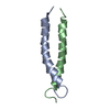
| |||||||||
|---|---|---|---|---|---|---|---|---|---|---|
| 1 |
| |||||||||
| NMR ensembles |
|
-Components
| #1: Protein/peptide | Mass: 4177.707 Da / Num. of mol.: 2 / Fragment: C-TERMINAL (RESIDUES 251-284) Source method: isolated from a genetically manipulated source Source: (gene. exp.)  Has protein modification | Y | |
|---|
-Experimental details
-Experiment
| Experiment | Method: SOLUTION NMR | ||||||||||||||||
|---|---|---|---|---|---|---|---|---|---|---|---|---|---|---|---|---|---|
| NMR experiment |
| ||||||||||||||||
| NMR details | Text: THE RESONANCE ASSIGNMENTS WERE MADE USING THROUGH BOND TRIPLE RESONANCE EXPERIMENTS INCLUDING HN(CA)CO, HNCO, H(CA)(C0)NH, H(CA)NH, CA(CO)NH, CANH, CBCA(CO)NH, CBCANH, HCCH-COSY AND CCH-COSY |
- Sample preparation
| Details | Contents: 1-2 MM Solvent system: 100 MM NACL, 20 MM POTASSIUM PHOSPHATE, 10% DUETERIUM OXIDE, 90% H20, PH 6.4 |
|---|---|
| Sample conditions | Ionic strength: 0.12 N / pH: 6.4 / Pressure: 1 atm / Temperature: 283.00 K |
| Crystal grow | *PLUS Method: other / Details: NMR |
-NMR measurement
| NMR spectrometer | Type: Varian INOVA / Manufacturer: Varian / Model: INOVA / Field strength: 600 MHz |
|---|
- Processing
| NMR software |
| ||||||||||||||||||||||||||||
|---|---|---|---|---|---|---|---|---|---|---|---|---|---|---|---|---|---|---|---|---|---|---|---|---|---|---|---|---|---|
| Refinement | Method: 1648 CONFORMATIONALLY- RESTRICTING NOE CONSTRAINTS (OUT OF A TOTAL OF 2036 NOE DERIVED DISTANCES) 52 LOOSE DIHEDRAL ANGLE CONSTRAINTS, 48 BACKBONE INTRA- HELICAL HYDROGEN BOND CONSTRAINTS WERE UTILILIZED. Software ordinal: 1 Details: REFINEMENT DETAILS CAN BE FOUND IN REFERENCE CITED ABOVE | ||||||||||||||||||||||||||||
| NMR representative | Selection criteria: fewest violations | ||||||||||||||||||||||||||||
| NMR ensemble | Conformer selection criteria: STRUCTURES WITH COVALENT GEOMETRY, STRUCTURES WITH THE LEAST RESTRAINT VIOLATIONS Conformers calculated total number: 200 / Conformers submitted total number: 10 |
