 Movie
Movie Controller


+ Open data

- Basic information
| Entry | Database: PDB / ID: 1mul | ||||||
|---|---|---|---|---|---|---|---|
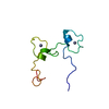
| Title | Crystal structure of the E. coli HU alpha2 protein | ||||||
 Components Components | DNA binding protein HU-alpha | ||||||
 Keywords Keywords | DNA BINDING PROTEIN / histone-like | ||||||
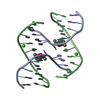
| Function / homology |  Function and homology information Function and homology informationHU-DNA complex / bacterial nucleoid DNA packaging / DnaA-HU complex / chromosome condensation / DNA replication initiation / structural constituent of chromatin / DNA repair / DNA damage response / DNA-templated transcription / DNA binding ...HU-DNA complex / bacterial nucleoid DNA packaging / DnaA-HU complex / chromosome condensation / DNA replication initiation / structural constituent of chromatin / DNA repair / DNA damage response / DNA-templated transcription / DNA binding / membrane / identical protein binding / cytosol Similarity search - Function | ||||||
| Biological species |  | ||||||
| Method |  X-RAY DIFFRACTION / MOLECULAR REPLACEMENT / Resolution: 2.3 Å X-RAY DIFFRACTION / MOLECULAR REPLACEMENT / Resolution: 2.3 Å | ||||||
 Authors Authors | Ramstein, J. / Hervouet, N. / Coste, F. / Zelwer, C. / Oberto, J. / Castaing, B. | ||||||
 Citation Citation | Journal: J.Mol.Biol. / Year: 2003 Title: Evidence of a Thermal Unfolding Dimeric Intermediate for the Escherichia coli Histone-like HU Proteins: Thermodynamics and Structure. Authors: Ramstein, J. / Hervouet, N. / Coste, F. / Zelwer, C. / Oberto, J. / Castaing, B. #1: Journal: Acta Crystallogr.,Sect.D / Year: 1999Title: Crystallization and preliminary X-ray diffraction analysis of the homodimeric form alpha2 of the HU protein from E. coli Authors: Coste, F. / Hervouet, N. / Oberto, J. / Zelwer, C. / Castaing, B. | ||||||
| History |
|
- Structure visualization
| Structure viewer | Molecule: MolmilJmol/JSmol |
|---|
- Downloads & links
-Download
| PDBx/mmCIF format | 1mul.cif.gz | 25.9 KB | Display | PDBx/mmCIF format |
|---|---|---|---|---|
| PDB format | pdb1mul.ent.gz | 16.7 KB | Display | PDB format |
| PDBx/mmJSON format | 1mul.json.gz | Tree view | PDBx/mmJSON format | |
| Others |  Other downloads Other downloads |
-Validation report
| Arichive directory | https://data.pdbj.org/pub/pdb/validation_reports/mu/1mulftp://data.pdbj.org/pub/pdb/validation_reports/mu/1mul | HTTPS FTP |
|---|
-Related structure data
| Similar structure data |
|---|


-Links
 PDBj
PDBj- Assembly
| Deposited unit | 
| ||||||||
|---|---|---|---|---|---|---|---|---|---|
| 1 |
| ||||||||
| 2 | 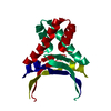
| ||||||||
| Unit cell |
|
-Components
| #1: Protein | Mass: 9549.979 Da / Num. of mol.: 1 Source method: isolated from a genetically manipulated source Source: (gene. exp.) |
|---|---|
| #2: Water | ChemComp-HOH /  Mass: 18.015 Da / Num. of mol.: 50 / Source method: isolated from a natural source / Formula: H2O Mass: 18.015 Da / Num. of mol.: 50 / Source method: isolated from a natural source / Formula: H2O |
-Experimental details
-Experiment
| Experiment | Method: X-RAY DIFFRACTION / Number of used crystals: 1 |
|---|
- Sample preparation
| Crystal | Density Matthews: 3.07 Å3/Da / Density % sol: 59.59 % |
|---|---|
| Crystal grow | Temperature: 293 K / Method: vapor diffusion, hanging drop / pH: 7.5 Details: PEG 4000, isopropanol, hepes, pH 7.5, VAPOR DIFFUSION, HANGING DROP, temperature 293K |
| Crystal grow | *PLUS Details: Coste, F., (1999) Acta Cryst., D55, 1952. |
-Data collection
| Diffraction | Mean temperature: 100 K |
|---|---|
| Diffraction source | Source: ROTATING ANODE / Type: RIGAKU RU200 / Wavelength: 1.5418 Å |
| Detector | Type: MARRESEARCH / Detector: IMAGE PLATE / Date: Jul 1, 1999 |
| Radiation | Monochromator: GRAPHITE / Protocol: SINGLE WAVELENGTH / Monochromatic (M) / Laue (L): M / Scattering type: x-ray |
| Radiation wavelength | Wavelength: 1.5418 Å / Relative weight: 1 |
| Reflection | Resolution: 2.3→32 Å / Num. all: 4755 / Num. obs: 4607 / Observed criterion σ(F): 1 / Observed criterion σ(I): 1 / Redundancy: 3.5 % / Biso Wilson estimate: 22.1 Å2 / Rsym value: 0.056 / Net I/σ(I): 11.2 |
| Reflection shell | Resolution: 2.3→2.36 Å / Redundancy: 3.6 % / Mean I/σ(I) obs: 4.4 / Num. unique all: 344 / Rsym value: 0.161 |
| Reflection | *PLUS Lowest resolution: 20 Å |
- Processing
| Software |
| ||||||||||||||||||||||||||||||||||||
|---|---|---|---|---|---|---|---|---|---|---|---|---|---|---|---|---|---|---|---|---|---|---|---|---|---|---|---|---|---|---|---|---|---|---|---|---|---|
| Refinement | Method to determine structure: MOLECULAR REPLACEMENT / Resolution: 2.3→20 Å / Rfactor Rfree error: 0.012 / Data cutoff high absF: 983947.18 / Data cutoff low absF: 0 / Isotropic thermal model: RESTRAINED / Cross valid method: THROUGHOUT / σ(F): 2 / Details: BULK SOLVENT MODEL USED
| ||||||||||||||||||||||||||||||||||||
| Solvent computation | Solvent model: FLAT MODEL / Bsol: 45.2065 Å2 / ksol: 0.358714 e/Å3 | ||||||||||||||||||||||||||||||||||||
| Displacement parameters | Biso mean: 27.9 Å2
| ||||||||||||||||||||||||||||||||||||
| Refine analyze |
| ||||||||||||||||||||||||||||||||||||
| Refinement step | Cycle: LAST / Resolution: 2.3→20 Å
| ||||||||||||||||||||||||||||||||||||
| Refine LS restraints |
| ||||||||||||||||||||||||||||||||||||
| LS refinement shell | Resolution: 2.3→2.44 Å / Rfactor Rfree error: 0.04 / Total num. of bins used: 6
| ||||||||||||||||||||||||||||||||||||
| Xplor file |
| ||||||||||||||||||||||||||||||||||||
| Refinement | *PLUS Highest resolution: 2.3 Å / Lowest resolution: 20 Å | ||||||||||||||||||||||||||||||||||||
| Solvent computation | *PLUS | ||||||||||||||||||||||||||||||||||||
| Displacement parameters | *PLUS | ||||||||||||||||||||||||||||||||||||
| Refine LS restraints | *PLUS
|
