 Movie
Movie Controller


+ Open data

- Basic information
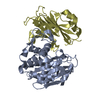
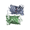
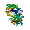
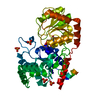
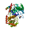
| Entry | Database: PDB / ID: 1msk | ||||||
|---|---|---|---|---|---|---|---|
| Title | METHIONINE SYNTHASE (ACTIVATION DOMAIN) | ||||||
 Components Components | COBALAMIN-DEPENDENT METHIONINE SYNTHASE | ||||||
 Keywords Keywords | METHYLTRANSFERASE / TRANSFERASE / METHIONINE BIOSYNTHESIS / VITAMIN B12 | ||||||
| Function / homology |  Function and homology information Function and homology informationmethionine synthase / methionine synthase activity / homocysteine metabolic process / methionine biosynthetic process / cobalamin binding / tetrahydrofolate metabolic process / tetrahydrofolate interconversion / methylation / zinc ion binding / cytoplasm / cytosol Similarity search - Function | ||||||
| Biological species |  | ||||||
| Method |  X-RAY DIFFRACTION / MIR / Resolution: 1.8 Å X-RAY DIFFRACTION / MIR / Resolution: 1.8 Å | ||||||
 Authors Authors | Dixon, M.M. / Huang, S. / Matthews, R.G. / Ludwig, M.L. | ||||||
 Citation Citation | Journal: Structure / Year: 1996 Title: The structure of the C-terminal domain of methionine synthase: presenting S-adenosylmethionine for reductive methylation of B12. Authors: Dixon, M.M. / Huang, S. / Matthews, R.G. / Ludwig, M. #1: Journal: Science / Year: 1994Title: How a Protein Binds B12: A 3.0 A X-Ray Structure of B12-Binding Domains of Methionine Synthase Authors: Drennan, C.L. / Huang, S. / Drummond, J.T. / Matthews, R.G. / Ludwig, M.L. | ||||||
| History |
|
- Structure visualization
| Structure viewer | Molecule: MolmilJmol/JSmol |
|---|
- Downloads & links
-Download
| PDBx/mmCIF format | 1msk.cif.gz | 84.3 KB | Display | PDBx/mmCIF format |
|---|---|---|---|---|
| PDB format | pdb1msk.ent.gz | 61.6 KB | Display | PDB format |
| PDBx/mmJSON format | 1msk.json.gz | Tree view | PDBx/mmJSON format | |
| Others |  Other downloads Other downloads |
-Validation report
| Summary document | 1msk_validation.pdf.gz | 446.6 KB | Display | wwPDB validaton report |
|---|---|---|---|---|
| Full document | 1msk_full_validation.pdf.gz | 451.4 KB | Display | |
| Data in XML | 1msk_validation.xml.gz | 8.6 KB | Display | |
| Data in CIF | 1msk_validation.cif.gz | 14 KB | Display | |
| Arichive directory | https://data.pdbj.org/pub/pdb/validation_reports/ms/1mskftp://data.pdbj.org/pub/pdb/validation_reports/ms/1msk | HTTPS FTP |
-Related structure data
| Similar structure data |
|---|


-Links
 PDBj
PDBj
- Assembly
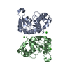
| Deposited unit | 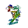
| ||||||||
|---|---|---|---|---|---|---|---|---|---|
| 1 |
| ||||||||
| Unit cell |
|
-Components
| #1: Protein | Mass: 37749.168 Da / Num. of mol.: 1 / Fragment: ACTIVATION DOMAIN, RESIDUES 897 - 1227 / Source method: isolated from a natural source Details: GENERATED BY TRYPSIN CLEAVAGE OF THE METHIONINE SYNTHASE HOLOENZYME Source: (natural) |
|---|---|
| #2: Chemical | ChemComp-ACT /   Mass: 59.044 Da / Num. of mol.: 1 / Source method: obtained synthetically / Formula: C2H3O2 Mass: 59.044 Da / Num. of mol.: 1 / Source method: obtained synthetically / Formula: C2H3O2 |
| #3: Chemical | ChemComp-SAM /   Mass: 398.437 Da / Num. of mol.: 1 / Source method: obtained synthetically / Formula: C15H22N6O5S Mass: 398.437 Da / Num. of mol.: 1 / Source method: obtained synthetically / Formula: C15H22N6O5S |
| #4: Water | ChemComp-HOH /  Mass: 18.015 Da / Num. of mol.: 212 / Source method: isolated from a natural source / Formula: H2O Mass: 18.015 Da / Num. of mol.: 212 / Source method: isolated from a natural source / Formula: H2O |
| Compound details | THE ACTIVATION |
-Experimental details
-Experiment
| Experiment | Method: X-RAY DIFFRACTION / Number of used crystals: 1 |
|---|
- Sample preparation
| Crystal | Density Matthews: 2.26 Å3/Da / Density % sol: 46 % | ||||||||||||||||||||||||||||||||||||||||||||||||
|---|---|---|---|---|---|---|---|---|---|---|---|---|---|---|---|---|---|---|---|---|---|---|---|---|---|---|---|---|---|---|---|---|---|---|---|---|---|---|---|---|---|---|---|---|---|---|---|---|---|
| Crystal grow | pH: 7.2 / Details: pH 7.2 | ||||||||||||||||||||||||||||||||||||||||||||||||
| Crystal grow | *PLUS Method: vapor diffusion, hanging drop | ||||||||||||||||||||||||||||||||||||||||||||||||
| Components of the solutions | *PLUS
|
-Data collection
| Diffraction | Mean temperature: 298 K |
|---|---|
| Diffraction source | Wavelength: 1.5418 |
| Detector | Type: ADSC / Detector: AREA DETECTOR |
| Radiation | Monochromatic (M) / Laue (L): M / Scattering type: x-ray |
| Radiation wavelength | Wavelength: 1.5418 Å / Relative weight: 1 |
| Reflection | Highest resolution: 1.8 Å / Num. obs: 28762 / % possible obs: 88 % / Observed criterion σ(I): 0 / Redundancy: 4.3 % / Rmerge(I) obs: 0.0532 / Net I/σ(I): 10.1 |
- Processing
| Software |
| ||||||||||||||||||||||||||||||||||||||||||||||||||||||||||||
|---|---|---|---|---|---|---|---|---|---|---|---|---|---|---|---|---|---|---|---|---|---|---|---|---|---|---|---|---|---|---|---|---|---|---|---|---|---|---|---|---|---|---|---|---|---|---|---|---|---|---|---|---|---|---|---|---|---|---|---|---|---|
| Refinement | Method to determine structure: MIR / Resolution: 1.8→10 Å / σ(F): 0 Details: TWO LOOPS, CONSISTING OF RESIDUES 965 - 967 AND 1035 - 1036, ARE DISORDERED, WITH AVERAGE MAIN-CHAIN B-FACTORS > 50.0 A**2. THREE RESIDUES AT THE C-TERMINAL END, 1225 - 1227, ARE DISORDERED, ...Details: TWO LOOPS, CONSISTING OF RESIDUES 965 - 967 AND 1035 - 1036, ARE DISORDERED, WITH AVERAGE MAIN-CHAIN B-FACTORS > 50.0 A**2. THREE RESIDUES AT THE C-TERMINAL END, 1225 - 1227, ARE DISORDERED, WITH AVERAGE MAIN-CHAIN B-FACTORS > 50.0 A**2.
| ||||||||||||||||||||||||||||||||||||||||||||||||||||||||||||
| Displacement parameters | Biso mean: 21.94 Å2 | ||||||||||||||||||||||||||||||||||||||||||||||||||||||||||||
| Refinement step | Cycle: LAST / Resolution: 1.8→10 Å
| ||||||||||||||||||||||||||||||||||||||||||||||||||||||||||||
| Refine LS restraints |
| ||||||||||||||||||||||||||||||||||||||||||||||||||||||||||||
| Software | *PLUS Name: X-PLOR / Classification: refinement | ||||||||||||||||||||||||||||||||||||||||||||||||||||||||||||
| Refinement | *PLUS | ||||||||||||||||||||||||||||||||||||||||||||||||||||||||||||
| Solvent computation | *PLUS | ||||||||||||||||||||||||||||||||||||||||||||||||||||||||||||
| Displacement parameters | *PLUS | ||||||||||||||||||||||||||||||||||||||||||||||||||||||||||||
| Refine LS restraints | *PLUS
|
