 Movie
Movie Controller

[English] 日本語
 Yorodumi
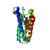
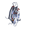
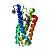
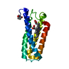
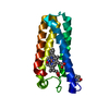
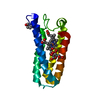
- PDB-1mqv: Crystal Structure of the Q1A/F32W/W72F mutant of Rhodopseudomonas... -
+ Open data

- Basic information
| Entry | Database: PDB / ID: 1mqv | |||||||||
|---|---|---|---|---|---|---|---|---|---|---|
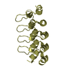
| Title | Crystal Structure of the Q1A/F32W/W72F mutant of Rhodopseudomonas palustris cytochrome c' (prime) expressed in E. coli | |||||||||
 Components Components | CYTOCHROME C' | |||||||||
 Keywords Keywords | ELECTRON TRANSPORT / Four-Helix Bundle | |||||||||
| Function / homology |  Function and homology information Function and homology informationelectron transport chain / periplasmic space / electron transfer activity / iron ion binding / heme binding Similarity search - Function | |||||||||
| Biological species |  Rhodopseudomonas palustris (phototrophic) Rhodopseudomonas palustris (phototrophic) | |||||||||
| Method |  X-RAY DIFFRACTION / MOLECULAR REPLACEMENT / Resolution: 1.78 Å X-RAY DIFFRACTION / MOLECULAR REPLACEMENT / Resolution: 1.78 Å | |||||||||
 Authors Authors | Lee, J.C. / Engman, K.C. / Tezcan, F.A. / Gray, H.B. / Winkler, J.R. | |||||||||
 Citation Citation | Journal: Proc.Natl.Acad.Sci.USA / Year: 2002 Title: Structural Features of Cytochrome c' Folding Intermediates Revealed by Fluorescence Energy-Transfer Kinetics Authors: Lee, J.C. / Engman, K.C. / Tezcan, F.A. / Gray, H.B. / Winkler, J.R. | |||||||||
| History |
|
- Structure visualization
| Structure viewer | Molecule: MolmilJmol/JSmol |
|---|
- Downloads & links
-Download
| PDBx/mmCIF format | 1mqv.cif.gz | 66.9 KB | Display | PDBx/mmCIF format |
|---|---|---|---|---|
| PDB format | pdb1mqv.ent.gz | 49.2 KB | Display | PDB format |
| PDBx/mmJSON format | 1mqv.json.gz | Tree view | PDBx/mmJSON format | |
| Others |  Other downloads Other downloads |
-Validation report
| Arichive directory | https://data.pdbj.org/pub/pdb/validation_reports/mq/1mqvftp://data.pdbj.org/pub/pdb/validation_reports/mq/1mqv | HTTPS FTP |
|---|
-Related structure data
| Related structure data | 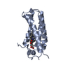 1a7vS S: Starting model for refinement |
|---|---|
| Similar structure data |

-Links
 PDBj
PDBj


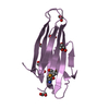
- Assembly
| Deposited unit | 
| ||||||||
|---|---|---|---|---|---|---|---|---|---|
| 1 | 
| ||||||||
| 2 | 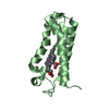
| ||||||||
| Unit cell |
|
-Components
| #1: Protein | Mass: 13095.089 Da / Num. of mol.: 2 / Mutation: Q1A, F32W, W72F Source method: isolated from a genetically manipulated source Details: The protein is in the OXIDIZED (FE(III)) STATE. Source: (gene. exp.) Rhodopseudomonas palustris (phototrophic)Plasmid: BL21(DE3) / Production host: #2: Chemical |   Mass: 618.503 Da / Num. of mol.: 2 / Source method: obtained synthetically / Formula: C34H34FeN4O4 Mass: 618.503 Da / Num. of mol.: 2 / Source method: obtained synthetically / Formula: C34H34FeN4O4#3: Water | ChemComp-HOH / |  Mass: 18.015 Da / Num. of mol.: 265 / Source method: isolated from a natural source / Formula: H2O Mass: 18.015 Da / Num. of mol.: 265 / Source method: isolated from a natural source / Formula: H2OHas protein modification | Y | |
|---|
-Experimental details
-Experiment
| Experiment | Method: X-RAY DIFFRACTION / Number of used crystals: 1 |
|---|
- Sample preparation
| Crystal | Density Matthews: 2.27 Å3/Da / Density % sol: 45.91 % | ||||||||||||||||||||||||||||||||||||||||||
|---|---|---|---|---|---|---|---|---|---|---|---|---|---|---|---|---|---|---|---|---|---|---|---|---|---|---|---|---|---|---|---|---|---|---|---|---|---|---|---|---|---|---|---|
| Crystal grow | Temperature: 298 K / Method: vapor diffusion, sitting drop / pH: 6 Details: PEG6000, sodium chloride, MES, pH 6.0, VAPOR DIFFUSION, SITTING DROP, temperature 298K | ||||||||||||||||||||||||||||||||||||||||||
| Crystal grow | *PLUS | ||||||||||||||||||||||||||||||||||||||||||
| Components of the solutions | *PLUS
|
-Data collection
| Diffraction | Mean temperature: 100 K |
|---|---|
| Diffraction source | Source: ROTATING ANODE / Type: RIGAKU RU200 / Wavelength: 1.5418 Å |
| Detector | Type: RIGAKU RAXIS II / Detector: IMAGE PLATE / Date: Mar 24, 2002 |
| Radiation | Monochromator: YALE MIRRORS / Protocol: SINGLE WAVELENGTH / Monochromatic (M) / Laue (L): M / Scattering type: x-ray |
| Radiation wavelength | Wavelength: 1.5418 Å / Relative weight: 1 |
| Reflection | Resolution: 1.78→50 Å / Num. all: 23685 / Num. obs: 23607 / % possible obs: 99.5 % / Observed criterion σ(F): 0 / Observed criterion σ(I): 0 / Rsym value: 0.062 / Net I/σ(I): 24.8 |
| Reflection shell | Resolution: 1.78→1.81 Å / Mean I/σ(I) obs: 1.9 / Rsym value: 0.487 / % possible all: 99 |
| Reflection | *PLUS Lowest resolution: 50 Å / % possible obs: 99.7 % / Rmerge(I) obs: 0.062 |
- Processing
| Software |
| |||||||||||||||||||||||||
|---|---|---|---|---|---|---|---|---|---|---|---|---|---|---|---|---|---|---|---|---|---|---|---|---|---|---|
| Refinement | Method to determine structure: MOLECULAR REPLACEMENT Starting model: PDB ENTRY 1A7V Resolution: 1.78→42 Å / Cross valid method: THROUGHOUT / σ(F): 0 / σ(I): 0 / Stereochemistry target values: Engh & Huber
| |||||||||||||||||||||||||
| Refine analyze |
| |||||||||||||||||||||||||
| Refinement step | Cycle: LAST / Resolution: 1.78→42 Å
| |||||||||||||||||||||||||
| Refine LS restraints |
| |||||||||||||||||||||||||
| LS refinement shell | Resolution: 1.78→1.84 Å / Rfactor Rfree error: 0.043
| |||||||||||||||||||||||||
| Refinement | *PLUS Lowest resolution: 42 Å / Rfactor Rwork: 0.211 | |||||||||||||||||||||||||
| Solvent computation | *PLUS | |||||||||||||||||||||||||
| Displacement parameters | *PLUS |
