 Movie
Movie Controller

[English] 日本語
 Yorodumi
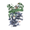
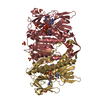
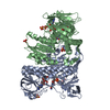
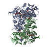
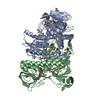
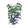
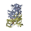
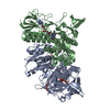
- PDB-1mok: NADPH DEPENDENT 2-KETOPROPYL COENZYME M OXIDOREDUCTASE/CARBOXYLASE -
+ Open data

- Basic information
| Entry | Database: PDB / ID: 1mok | ||||||
|---|---|---|---|---|---|---|---|
| Title | NADPH DEPENDENT 2-KETOPROPYL COENZYME M OXIDOREDUCTASE/CARBOXYLASE | ||||||
 Components Components | orf3 | ||||||
 Keywords Keywords | OXIDOREDUCTASE / Nucleotide binding motifs / Nucleotide binding domain | ||||||
| Function / homology |  Function and homology information Function and homology information2-oxopropyl-CoM reductase (carboxylating) / 2-oxopropyl-CoM reductase (carboxylating) activity / propylene catabolic process Similarity search - Function | ||||||
| Biological species |  Xanthobacter autotrophicus (bacteria) Xanthobacter autotrophicus (bacteria) | ||||||
| Method |  X-RAY DIFFRACTION / SYNCHROTRON / MOLECULAR REPLACEMENT / Resolution: 2.8 Å X-RAY DIFFRACTION / SYNCHROTRON / MOLECULAR REPLACEMENT / Resolution: 2.8 Å | ||||||
 Authors Authors | Nocek, B. / Jang, S.B. / Jeong, M.S. / Clark, D.D. / Ensign, S.A. / Peters, J.W. | ||||||
 Citation Citation | Journal: Biochemistry / Year: 2002 Title: Structural Basis for CO2 Fixation by a Novel Member of the Disulfide Oxidoreductase Family of Enzymes, 2-Ketopropyl Coenzyme M Oxidoreductase/Carboxylase Authors: Nocek, B. / Jang, S.B. / Jeong, M.S. / Clark, D.D. / Ensign, S.A. / Peters, J.W. | ||||||
| History |
|
- Structure visualization
| Structure viewer | Molecule: MolmilJmol/JSmol |
|---|
- Downloads & links
-Download
| PDBx/mmCIF format | 1mok.cif.gz | 398.8 KB | Display | PDBx/mmCIF format |
|---|---|---|---|---|
| PDB format | pdb1mok.ent.gz | 326.7 KB | Display | PDB format |
| PDBx/mmJSON format | 1mok.json.gz | Tree view | PDBx/mmJSON format | |
| Others |  Other downloads Other downloads |
-Validation report
| Summary document | 1mok_validation.pdf.gz | 685.3 KB | Display | wwPDB validaton report |
|---|---|---|---|---|
| Full document | 1mok_full_validation.pdf.gz | 775.5 KB | Display | |
| Data in XML | 1mok_validation.xml.gz | 53.9 KB | Display | |
| Data in CIF | 1mok_validation.cif.gz | 76.9 KB | Display | |
| Arichive directory | https://data.pdbj.org/pub/pdb/validation_reports/mo/1mokftp://data.pdbj.org/pub/pdb/validation_reports/mo/1mok | HTTPS FTP |
-Related structure data
| Related structure data |  1mo9SC S: Starting model for refinement C: citing same article ( |
|---|---|
| Similar structure data |


-Links
 PDBj
PDBj


- Assembly
| Deposited unit | 
| ||||||||
|---|---|---|---|---|---|---|---|---|---|
| 1 | 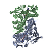
| ||||||||
| 2 | 
| ||||||||
| Unit cell |
|
-Components
| #1: Protein | Mass: 57414.348 Da / Num. of mol.: 4 / Source method: isolated from a natural source / Source: (natural) Xanthobacter autotrophicus (bacteria) / Strain: Py2References: UniProt: Q56839, 2-oxopropyl-CoM reductase (carboxylating) #2: Chemical | ChemComp-FAD /   Mass: 785.550 Da / Num. of mol.: 4 / Source method: obtained synthetically / Formula: C27H33N9O15P2 / Comment: FAD*YM Mass: 785.550 Da / Num. of mol.: 4 / Source method: obtained synthetically / Formula: C27H33N9O15P2 / Comment: FAD*YMHas protein modification | Y | |
|---|
-Experimental details
-Experiment
| Experiment | Method: X-RAY DIFFRACTION / Number of used crystals: 1 |
|---|
- Sample preparation
| Crystal | Density Matthews: 2.2 Å3/Da / Density % sol: 44.09 % | ||||||||||||||||||||||||||||||||||||||||||
|---|---|---|---|---|---|---|---|---|---|---|---|---|---|---|---|---|---|---|---|---|---|---|---|---|---|---|---|---|---|---|---|---|---|---|---|---|---|---|---|---|---|---|---|
| Crystal grow | Temperature: 294 K / Method: vapor diffusion, hanging drop / pH: 6.5 Details: 0.17M ammonium acetate, 0.085M Tris-HCl pH 8.5, 25.5% PEG 4000, 15% glycerol, pH 6.5, VAPOR DIFFUSION, HANGING DROP, temperature 294K | ||||||||||||||||||||||||||||||||||||||||||
| Crystal grow | *PLUS pH: 5.6 / Details: Jang, S.B., (2001) Acta Crystallogr., D57, 445. | ||||||||||||||||||||||||||||||||||||||||||
| Components of the solutions | *PLUS
|
-Data collection
| Diffraction | Mean temperature: 93 K |
|---|---|
| Diffraction source | Source: SYNCHROTRON / Site: SSRL  / Beamline: BL9-1 / Wavelength: 0.78 Å / Beamline: BL9-1 / Wavelength: 0.78 Å |
| Detector | Type: MAR scanner 345 mm plate / Detector: IMAGE PLATE / Date: Feb 19, 2000 / Details: flat mirrors |
| Radiation | Protocol: SINGLE WAVELENGTH / Monochromatic (M) / Laue (L): M / Scattering type: x-ray |
| Radiation wavelength | Wavelength: 0.78 Å / Relative weight: 1 |
| Reflection | Resolution: 2.8→20 Å / Num. all: 41494 / Num. obs: 41494 / Observed criterion σ(I): 0.5 |
| Reflection | *PLUS Num. obs: 48234 / % possible obs: 92.1 % / Num. measured all: 213523 / Rmerge(I) obs: 0.098 |
- Processing
| Software |
| |||||||||||||||||||||||||||
|---|---|---|---|---|---|---|---|---|---|---|---|---|---|---|---|---|---|---|---|---|---|---|---|---|---|---|---|---|
| Refinement | Method to determine structure: MOLECULAR REPLACEMENT Starting model: PDB ENTRY 1MO9 Resolution: 2.8→19.99 Å / Cross valid method: THROUGHOUT / σ(F): 0.5 / Stereochemistry target values: Engh & Huber
| |||||||||||||||||||||||||||
| Displacement parameters | Biso mean: 35.2 Å2
| |||||||||||||||||||||||||||
| Refinement step | Cycle: LAST / Resolution: 2.8→19.99 Å
| |||||||||||||||||||||||||||
| Refinement | *PLUS Lowest resolution: 20 Å / Rfactor Rfree: 0.276 / Rfactor Rwork: 0.224 | |||||||||||||||||||||||||||
| Solvent computation | *PLUS | |||||||||||||||||||||||||||
| Displacement parameters | *PLUS | |||||||||||||||||||||||||||
| Refine LS restraints | *PLUS
|
