 Movie
Movie Controller

[English] 日本語
 Yorodumi
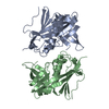
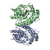
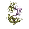
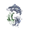
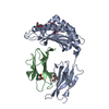
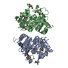
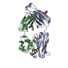
- PDB-1moe: The three-dimensional structure of an engineered scFv T84.66 dime... -
+ Open data

- Basic information
| Entry | Database: PDB / ID: 1moe | ||||||
|---|---|---|---|---|---|---|---|
| Title | The three-dimensional structure of an engineered scFv T84.66 dimer or diabody in VL to VH linkage. | ||||||
 Components Components | anti-CEA mAb T84.66 | ||||||
 Keywords Keywords | IMMUNE SYSTEM / anti carcinoembryonic antigen / T84.66 / diabody / dimer / scFv / variable domain | ||||||
| Function / homology |  Function and homology information Function and homology informationimmunoglobulin complex / adaptive immune response / immune response / extracellular region / plasma membrane Similarity search - Function | ||||||
| Biological species |  | ||||||
| Method |  X-RAY DIFFRACTION / MOLECULAR REPLACEMENT / Resolution: 2.6 Å X-RAY DIFFRACTION / MOLECULAR REPLACEMENT / Resolution: 2.6 Å | ||||||
 Authors Authors | Carmichael, J.A. / Power, B.E. / Garrett, T.P.J. / Yazaki, P.J. / Shively, J.E. / Raubischek, A.A. / Wu, A.M. / Hudson, P.J. | ||||||
 Citation Citation | Journal: J.Mol.Biol. / Year: 2003 Title: The Crystal Structure of an Anti-CEA scFv Diabody Assembled from T84.66 scFvs in VL-to-VH Orientation: Implications for Diabody Flexibility Authors: Carmichael, J.A. / Power, B.E. / Garrett, T.P. / Yazaki, P.J. / Shively, J.E. / Raubischek, A.A. / Wu, A.M. / Hudson, P.J. #1: Journal: J.Immunol.Methods / Year: 2001Title: Mammalian expression and hollow fiber bioreactor production of recombinant anti-CEA diabody and minibody for clinical applications. Authors: Yazaki, P.J. / Shively, L. / Clark, C. / Cheung, C.W. / Le, W. / Szpikowska, B. / Shively, J.E. / Raubitschek, A.A. / Wu, A.M. #2: Journal: Acta Crystallogr.,Sect.A / Year: 1999Title: Structure of an Exoglucanase complexed with conduritol B epoxide from A 50um Crystal using monocappilary optics. Authors: Varghese, L.N. / Van Donkelaar, A. / Hrmova, M. / Fincher, G.A. / Baliac, D.X. / Barnia, Z. | ||||||
| History |
|
- Structure visualization
| Structure viewer | Molecule: MolmilJmol/JSmol |
|---|
- Downloads & links
-Download
| PDBx/mmCIF format | 1moe.cif.gz | 104.3 KB | Display | PDBx/mmCIF format |
|---|---|---|---|---|
| PDB format | pdb1moe.ent.gz | 81 KB | Display | PDB format |
| PDBx/mmJSON format | 1moe.json.gz | Tree view | PDBx/mmJSON format | |
| Others |  Other downloads Other downloads |
-Validation report
| Arichive directory | https://data.pdbj.org/pub/pdb/validation_reports/mo/1moeftp://data.pdbj.org/pub/pdb/validation_reports/mo/1moe | HTTPS FTP |
|---|
-Related structure data
| Related structure data | 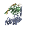 1gg2S S: Starting model for refinement |
|---|---|
| Similar structure data |


-Links
 PDBj
PDBj

- Assembly
| Deposited unit | 
| ||||||||
|---|---|---|---|---|---|---|---|---|---|
| 1 |
| ||||||||
| 2 | 
| ||||||||
| Unit cell |
|
-Components
| #1: Antibody | Mass: 25815.623 Da / Num. of mol.: 2 / Fragment: RESIDUES 1-240 Source method: isolated from a genetically manipulated source Details: immunoglobulin variable domains linked VL to VH with (GGGSGGGG) Source: (gene. exp.) References: UniProt: P01660, GenBank: 50373, UniProt: Q5R3X1*PLUS #2: Chemical |   Mass: 96.063 Da / Num. of mol.: 3 / Source method: obtained synthetically / Formula: SO4 Mass: 96.063 Da / Num. of mol.: 3 / Source method: obtained synthetically / Formula: SO4#3: Water | ChemComp-HOH / |  Mass: 18.015 Da / Num. of mol.: 71 / Source method: isolated from a natural source / Formula: H2O Mass: 18.015 Da / Num. of mol.: 71 / Source method: isolated from a natural source / Formula: H2OHas protein modification | Y | |
|---|
-Experimental details
-Experiment
| Experiment | Method: X-RAY DIFFRACTION / Number of used crystals: 2 |
|---|
- Sample preparation
| Crystal | Density Matthews: 2.46 Å3/Da / Density % sol: 49.61 % | |||||||||||||||||||||||||||||||||||
|---|---|---|---|---|---|---|---|---|---|---|---|---|---|---|---|---|---|---|---|---|---|---|---|---|---|---|---|---|---|---|---|---|---|---|---|---|
| Crystal grow | Temperature: 294 K / Method: vapor diffusion, hanging drop / pH: 7.4 Details: sodium chloride, HEPES, ammonium sulfate, pH 7.4, VAPOR DIFFUSION, HANGING DROP, temperature 294K | |||||||||||||||||||||||||||||||||||
| Crystal grow | *PLUS pH: 7.5 | |||||||||||||||||||||||||||||||||||
| Components of the solutions | *PLUS
|
-Data collection
| Diffraction |
| ||||||||||||||||||
|---|---|---|---|---|---|---|---|---|---|---|---|---|---|---|---|---|---|---|---|
| Diffraction source | Source: ROTATING ANODE / Type: RIGAKU / Wavelength: 1.5418 Å | ||||||||||||||||||
| Detector |
| ||||||||||||||||||
| Radiation |
| ||||||||||||||||||
| Radiation wavelength | Wavelength: 1.5418 Å / Relative weight: 1 | ||||||||||||||||||
| Reflection | Resolution: 2.5→20 Å / Num. all: 19293 / Num. obs: 17303 / % possible obs: 90 % / Observed criterion σ(F): 2 / Observed criterion σ(I): 1 / Redundancy: 5.3 % / Biso Wilson estimate: 14.2 Å2 / Limit h max: 24 / Limit h min: 0 / Limit k max: 18 / Limit k min: 0 / Limit l max: 96 / Limit l min: 0 / Observed criterion F max: 322511.49 / Observed criterion F min: 0.32 / Rmerge(I) obs: 0.072 / Net I/σ(I): 18.8 | ||||||||||||||||||
| Reflection shell | Resolution: 2.5→2.59 Å / Redundancy: 2.4 % / Rmerge(I) obs: 0.439 / Mean I/σ(I) obs: 1.5 / Num. unique all: 1347 / % possible all: 72 | ||||||||||||||||||
| Reflection | *PLUS Lowest resolution: 20 Å / Num. obs: 15066 / % possible obs: 86.2 % / Redundancy: 52.4 % / Rmerge(I) obs: 0.075 | ||||||||||||||||||
| Reflection shell | *PLUS % possible obs: 44.2 % / Redundancy: 18.5 % / Num. unique obs: 819 / Rmerge(I) obs: 0.28 / Mean I/σ(I) obs: 3.5 |
- Processing
| Software |
| ||||||||||||||||||||||||||||||||||||||||||||||||||||||||||||||||||||||||||||||||||||||||||||||||||||||||||||||
|---|---|---|---|---|---|---|---|---|---|---|---|---|---|---|---|---|---|---|---|---|---|---|---|---|---|---|---|---|---|---|---|---|---|---|---|---|---|---|---|---|---|---|---|---|---|---|---|---|---|---|---|---|---|---|---|---|---|---|---|---|---|---|---|---|---|---|---|---|---|---|---|---|---|---|---|---|---|---|---|---|---|---|---|---|---|---|---|---|---|---|---|---|---|---|---|---|---|---|---|---|---|---|---|---|---|---|---|---|---|---|---|
| Refinement | Method to determine structure: MOLECULAR REPLACEMENT Starting model: 1gg2 Resolution: 2.6→20 Å / Rfactor Rfree error: 0.009 / Occupancy max: 1 / Occupancy min: 1 / Isotropic thermal model: anisotropic / Cross valid method: THROUGHOUT / σ(F): 2 / σ(I): 2 / Stereochemistry target values: Engh & Huber
| ||||||||||||||||||||||||||||||||||||||||||||||||||||||||||||||||||||||||||||||||||||||||||||||||||||||||||||||
| Solvent computation | Solvent model: CNS bulk solvent model used / Bsol: 35.2171 Å2 / ksol: 0.30081 e/Å3 | ||||||||||||||||||||||||||||||||||||||||||||||||||||||||||||||||||||||||||||||||||||||||||||||||||||||||||||||
| Displacement parameters | Biso max: 104.15 Å2 / Biso mean: 42.58 Å2 / Biso min: 10.7 Å2
| ||||||||||||||||||||||||||||||||||||||||||||||||||||||||||||||||||||||||||||||||||||||||||||||||||||||||||||||
| Refine analyze |
| ||||||||||||||||||||||||||||||||||||||||||||||||||||||||||||||||||||||||||||||||||||||||||||||||||||||||||||||
| Refinement step | Cycle: LAST / Resolution: 2.6→20 Å
| ||||||||||||||||||||||||||||||||||||||||||||||||||||||||||||||||||||||||||||||||||||||||||||||||||||||||||||||
| Refine LS restraints |
| ||||||||||||||||||||||||||||||||||||||||||||||||||||||||||||||||||||||||||||||||||||||||||||||||||||||||||||||
| LS refinement shell | Refine-ID: X-RAY DIFFRACTION / Total num. of bins used: 10
| ||||||||||||||||||||||||||||||||||||||||||||||||||||||||||||||||||||||||||||||||||||||||||||||||||||||||||||||
| Xplor file |
| ||||||||||||||||||||||||||||||||||||||||||||||||||||||||||||||||||||||||||||||||||||||||||||||||||||||||||||||
| Software | *PLUS Name: CNS / Classification: refinement | ||||||||||||||||||||||||||||||||||||||||||||||||||||||||||||||||||||||||||||||||||||||||||||||||||||||||||||||
| Refinement | *PLUS Lowest resolution: 20 Å / % reflection Rfree: 10 % / Rfactor Rfree: 0.298 / Rfactor Rwork: 0.215 | ||||||||||||||||||||||||||||||||||||||||||||||||||||||||||||||||||||||||||||||||||||||||||||||||||||||||||||||
| Solvent computation | *PLUS | ||||||||||||||||||||||||||||||||||||||||||||||||||||||||||||||||||||||||||||||||||||||||||||||||||||||||||||||
| Displacement parameters | *PLUS | ||||||||||||||||||||||||||||||||||||||||||||||||||||||||||||||||||||||||||||||||||||||||||||||||||||||||||||||
| Refine LS restraints | *PLUS
|
