 Movie
Movie Controller

[English] 日本語
 Yorodumi
- PDB-1mo0: Structural Genomics Of Caenorhabditis Elegans: Triose Phosphate I... -
+ Open data

- Basic information
| Entry | Database: PDB / ID: 1mo0 | ||||||
|---|---|---|---|---|---|---|---|
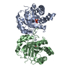
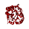
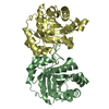
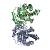
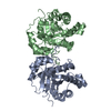
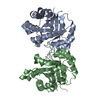
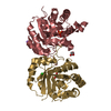
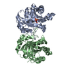
| Title | Structural Genomics Of Caenorhabditis Elegans: Triose Phosphate Isomerase | ||||||
 Components Components | Triosephosphate isomerase | ||||||
 Keywords Keywords | ISOMERASE / STRUCTURAL GENOMICS / Triose Phosphate Isomerase / PSI / Protein Structure Initiative / Southeast Collaboratory for Structural Genomics / SECSG | ||||||
| Function / homology |  Function and homology information Function and homology informationGlycolysis / Gluconeogenesis / methylglyoxal synthase / methylglyoxal synthase activity / triose-phosphate isomerase / triose-phosphate isomerase activity / glyceraldehyde-3-phosphate biosynthetic process / glycerol catabolic process / myofibril / cell redox homeostasis ...Glycolysis / Gluconeogenesis / methylglyoxal synthase / methylglyoxal synthase activity / triose-phosphate isomerase / triose-phosphate isomerase activity / glyceraldehyde-3-phosphate biosynthetic process / glycerol catabolic process / myofibril / cell redox homeostasis / glycolytic process / determination of adult lifespan / gluconeogenesis / cytosol Similarity search - Function | ||||||
| Biological species |  | ||||||
| Method |  X-RAY DIFFRACTION / SYNCHROTRON / SAS / Resolution: 1.7 Å X-RAY DIFFRACTION / SYNCHROTRON / SAS / Resolution: 1.7 Å | ||||||
 Authors Authors | Symersky, J. / Li, S. / Finley, J. / Liu, Z.-J. / Qui, H. / Luan, C.H. / Carson, M. / Tsao, J. / Johnson, D. / Lin, G. ...Symersky, J. / Li, S. / Finley, J. / Liu, Z.-J. / Qui, H. / Luan, C.H. / Carson, M. / Tsao, J. / Johnson, D. / Lin, G. / Zhao, J. / Thomas, W. / Nagy, L.A. / Sha, B. / DeLucas, L.J. / Wang, B.-C. / Luo, M. / Southeast Collaboratory for Structural Genomics (SECSG) | ||||||
 Citation Citation | Journal: Proteins / Year: 2003 Title: Structural genomics of Caenorhabditis elegans: triosephosphate isomerase Authors: Symersky, J. / Li, S. / Carson, M. / Luo, M. #1: Journal: To be publishedTitle: Structural genomics of Caenorhabditis elegans: The structure of the cytoskeleton-associated protein (CAP-Gly) domain of F53F4.3. Authors: Li, S. / Finley, J. / Liu, Z.-J. / Qui, H. / Luan, C.H. / Carson, M. / Tsao, J. / Johnson, D. / Lin, G. / Zhao, J. / Thomas, W. / Nagy, L.A. / Sha, B. / DeLucas, L.J. / Wang, B.-C. / Luo, M. | ||||||
| History |
|
- Structure visualization
| Structure viewer | Molecule: MolmilJmol/JSmol |
|---|
- Downloads & links
-Download
| PDBx/mmCIF format | 1mo0.cif.gz | 117.9 KB | Display | PDBx/mmCIF format |
|---|---|---|---|---|
| PDB format | pdb1mo0.ent.gz | 89.9 KB | Display | PDB format |
| PDBx/mmJSON format | 1mo0.json.gz | Tree view | PDBx/mmJSON format | |
| Others |  Other downloads Other downloads |
-Validation report
| Arichive directory | https://data.pdbj.org/pub/pdb/validation_reports/mo/1mo0ftp://data.pdbj.org/pub/pdb/validation_reports/mo/1mo0 | HTTPS FTP |
|---|
-Related structure data
| Related structure data | |
|---|---|
| Similar structure data | |
| Other databases |


-Links
 PDBj
PDBj
- Assembly
| Deposited unit | 
| ||||||||||
|---|---|---|---|---|---|---|---|---|---|---|---|
| 1 |
| ||||||||||
| Unit cell |
|
-Components
| #1: Protein | Mass: 29880.043 Da / Num. of mol.: 2 Source method: isolated from a genetically manipulated source Source: (gene. exp.)  #2: Chemical |   Mass: 59.044 Da / Num. of mol.: 2 / Source method: obtained synthetically / Formula: C2H3O2 Mass: 59.044 Da / Num. of mol.: 2 / Source method: obtained synthetically / Formula: C2H3O2#3: Chemical | ChemComp-SO4 /   Mass: 96.063 Da / Num. of mol.: 5 / Source method: obtained synthetically / Formula: SO4 Mass: 96.063 Da / Num. of mol.: 5 / Source method: obtained synthetically / Formula: SO4#4: Water | ChemComp-HOH / |  Mass: 18.015 Da / Num. of mol.: 461 / Source method: isolated from a natural source / Formula: H2O Mass: 18.015 Da / Num. of mol.: 461 / Source method: isolated from a natural source / Formula: H2O |
|---|
-Experimental details
-Experiment
| Experiment | Method: X-RAY DIFFRACTION / Number of used crystals: 1 |
|---|
- Sample preparation
| Crystal | Density Matthews: 2.063 Å3/Da / Density % sol: 38.09 % | ||||||||||||||||||||||||||||||
|---|---|---|---|---|---|---|---|---|---|---|---|---|---|---|---|---|---|---|---|---|---|---|---|---|---|---|---|---|---|---|---|
| Crystal grow | Temperature: 293 K / Method: vapor diffusion, hanging drop / pH: 5.5 Details: 2 M AMMONIUM SULFATE, 50 mM SODIUM ACETATE at pH 5.5, VAPOR DIFFUSION, HANGING DROP, temperature 293K | ||||||||||||||||||||||||||||||
| Crystal grow | *PLUS Temperature: 22 ℃ / pH: 7.5 | ||||||||||||||||||||||||||||||
| Components of the solutions | *PLUS
|
-Data collection
| Diffraction | Mean temperature: 100 K |
|---|---|
| Diffraction source | Source: SYNCHROTRON / Site: SSRL  / Beamline: BL9-1 / Wavelength: 0.976 / Beamline: BL9-1 / Wavelength: 0.976 |
| Detector | Detector: IMAGE PLATE / Date: Jun 22, 2002 |
| Radiation | Protocol: SINGLE WAVELENGTH / Monochromatic (M) / Laue (L): M / Scattering type: x-ray |
| Radiation wavelength | Wavelength: 0.976 Å / Relative weight: 1 |
| Reflection | Resolution: 1.7→29.6 Å / Num. all: 49911 / Num. obs: 49911 / % possible obs: 92.7 % / Observed criterion σ(I): 0 / Redundancy: 3.49 % / Biso Wilson estimate: 14.6 Å2 / Net I/σ(I): 15.1 |
| Reflection shell | Resolution: 1.7→1.76 Å / % possible obs: 86.5 % / Redundancy: 2.64 % / Rsym value: 0.206 |
| Reflection | *PLUS Highest resolution: 1.7 Å / Lowest resolution: 29.6 Å / Num. obs: 49994 / Num. measured all: 174695 / Rmerge(I) obs: 0.039 |
| Reflection shell | *PLUS Highest resolution: 1.7 Å / Rmerge(I) obs: 0.206 |
- Processing
| Software |
| ||||||||||||||||||||||||||||||||||||||||||||||||||||||||||||||||||||||||||||||||
|---|---|---|---|---|---|---|---|---|---|---|---|---|---|---|---|---|---|---|---|---|---|---|---|---|---|---|---|---|---|---|---|---|---|---|---|---|---|---|---|---|---|---|---|---|---|---|---|---|---|---|---|---|---|---|---|---|---|---|---|---|---|---|---|---|---|---|---|---|---|---|---|---|---|---|---|---|---|---|---|---|---|
| Refinement | Method to determine structure: SAS Starting model: THE STRUCTURE WAS SOLVED USING THE DATA COLLECTED ON R-AXIS IV WITH CU_KA RADIATION TO 2-A RESOLUTION. THE CRYSTAL WAS SOAKED WITH POTASSIUM IODIDE PRIOR DATA COLLECTION Resolution: 1.7→29.6 Å / Rfactor Rfree error: 0.004 / Data cutoff high absF: 1000 / Data cutoff low absF: 0 / Isotropic thermal model: RESTRAINED / Cross valid method: THROUGHOUT / σ(F): 0 / Stereochemistry target values: MLF
| ||||||||||||||||||||||||||||||||||||||||||||||||||||||||||||||||||||||||||||||||
| Solvent computation | Solvent model: FLAT MODEL / Bsol: 53.8849 Å2 / ksol: 0.384014 e/Å3 | ||||||||||||||||||||||||||||||||||||||||||||||||||||||||||||||||||||||||||||||||
| Displacement parameters | Biso mean: 16.5 Å2
| ||||||||||||||||||||||||||||||||||||||||||||||||||||||||||||||||||||||||||||||||
| Refine analyze | Luzzati coordinate error free: 0.21 Å / Luzzati sigma a free: 0.19 Å | ||||||||||||||||||||||||||||||||||||||||||||||||||||||||||||||||||||||||||||||||
| Refinement step | Cycle: LAST / Resolution: 1.7→29.6 Å
| ||||||||||||||||||||||||||||||||||||||||||||||||||||||||||||||||||||||||||||||||
| Refine LS restraints |
| ||||||||||||||||||||||||||||||||||||||||||||||||||||||||||||||||||||||||||||||||
| LS refinement shell | Resolution: 1.7→1.81 Å / Rfactor Rfree error: 0.014 / Total num. of bins used: 6
| ||||||||||||||||||||||||||||||||||||||||||||||||||||||||||||||||||||||||||||||||
| Xplor file |
| ||||||||||||||||||||||||||||||||||||||||||||||||||||||||||||||||||||||||||||||||
| Refinement | *PLUS Highest resolution: 1.7 Å / Lowest resolution: 29.6 Å / Num. reflection obs: 49994 / % reflection Rfree: 5 % | ||||||||||||||||||||||||||||||||||||||||||||||||||||||||||||||||||||||||||||||||
| Solvent computation | *PLUS | ||||||||||||||||||||||||||||||||||||||||||||||||||||||||||||||||||||||||||||||||
| Displacement parameters | *PLUS | ||||||||||||||||||||||||||||||||||||||||||||||||||||||||||||||||||||||||||||||||
| Refine LS restraints | *PLUS
|
