 Movie
Movie Controller

[English] 日本語
 Yorodumi
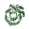
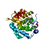
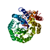
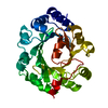
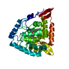
- PDB-1mlw: Crystal structure of human tryptophan hydroxylase with bound 7,8-... -
+ Open data

- Basic information
| Entry | Database: PDB / ID: 1mlw | ||||||
|---|---|---|---|---|---|---|---|
| Title | Crystal structure of human tryptophan hydroxylase with bound 7,8-dihydro-L-biopterin cofactor and Fe(III) | ||||||
 Components Components | Tryptophan 5-monooxygenase | ||||||
 Keywords Keywords | OXIDOREDUCTASE / aromatic amino acid hydroxylase catalytic domain fold | ||||||
| Function / homology |  Function and homology information Function and homology informationregulation of hemostasis / : / tryptophan 5-monooxygenase / tryptophan 5-monooxygenase activity / Serotonin and melatonin biosynthesis / serotonin biosynthetic process / platelet degranulation / bone remodeling / NGF-stimulated transcription / mammary gland alveolus development ...regulation of hemostasis / : / tryptophan 5-monooxygenase / tryptophan 5-monooxygenase activity / Serotonin and melatonin biosynthesis / serotonin biosynthetic process / platelet degranulation / bone remodeling / NGF-stimulated transcription / mammary gland alveolus development / positive regulation of fat cell differentiation / neuron projection / iron ion binding / cytosol Similarity search - Function | ||||||
| Biological species |  Homo sapiens (human) Homo sapiens (human) | ||||||
| Method |  X-RAY DIFFRACTION / SYNCHROTRON / MOLECULAR REPLACEMENT / Resolution: 1.71 Å X-RAY DIFFRACTION / SYNCHROTRON / MOLECULAR REPLACEMENT / Resolution: 1.71 Å | ||||||
 Authors Authors | Wang, L. / Erlandsen, H. / Haavik, J. / Knappskog, P.M. / Stevens, R.C. | ||||||
 Citation Citation | Journal: Biochemistry / Year: 2002 Title: Three-dimensional structure of human tryptophan hydroxylase and its implications for the biosynthesis of the neurotransmitters serotonin and melatonin Authors: Wang, L. / Erlandsen, H. / Haavik, J. / Knappskog, P.M. / Stevens, R.C. | ||||||
| History |
|
- Structure visualization
| Structure viewer | Molecule: MolmilJmol/JSmol |
|---|
- Downloads & links
-Download
| PDBx/mmCIF format | 1mlw.cif.gz | 75.8 KB | Display | PDBx/mmCIF format |
|---|---|---|---|---|
| PDB format | pdb1mlw.ent.gz | 56.1 KB | Display | PDB format |
| PDBx/mmJSON format | 1mlw.json.gz | Tree view | PDBx/mmJSON format | |
| Others |  Other downloads Other downloads |
-Validation report
| Arichive directory | https://data.pdbj.org/pub/pdb/validation_reports/ml/1mlwftp://data.pdbj.org/pub/pdb/validation_reports/ml/1mlw | HTTPS FTP |
|---|
-Related structure data
| Related structure data |  1pahS S: Starting model for refinement |
|---|---|
| Similar structure data |


-Links
 PDBj
PDBj


- Assembly
| Deposited unit | 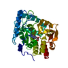
| ||||||||
|---|---|---|---|---|---|---|---|---|---|
| 1 |
| ||||||||
| Unit cell |
| ||||||||
| Details | biological unit is a monomer |
-Components
| #1: Protein | Mass: 34641.523 Da / Num. of mol.: 1 Fragment: Double truncated tryptophan hydroxylase catalytic domain (Residues 102-402) Source method: isolated from a genetically manipulated source Source: (gene. exp.) Homo sapiens (human) / Gene: TPH / Plasmid: pET23a / Production host:  |
|---|---|
| #2: Chemical | ChemComp-FE /   Mass: 55.845 Da / Num. of mol.: 1 / Source method: obtained synthetically / Formula: Fe Mass: 55.845 Da / Num. of mol.: 1 / Source method: obtained synthetically / Formula: Fe |
| #3: Chemical | ChemComp-HBI / 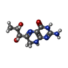  Mass: 239.231 Da / Num. of mol.: 1 / Source method: obtained synthetically / Formula: C9H13N5O3 Mass: 239.231 Da / Num. of mol.: 1 / Source method: obtained synthetically / Formula: C9H13N5O3 |
| #4: Water | ChemComp-HOH /  Mass: 18.015 Da / Num. of mol.: 242 / Source method: isolated from a natural source / Formula: H2O Mass: 18.015 Da / Num. of mol.: 242 / Source method: isolated from a natural source / Formula: H2O |
-Experimental details
-Experiment
| Experiment | Method: X-RAY DIFFRACTION / Number of used crystals: 1 |
|---|
- Sample preparation
| Crystal | Density Matthews: 2.16 Å3/Da / Density % sol: 43.1 % | ||||||||||||||||||
|---|---|---|---|---|---|---|---|---|---|---|---|---|---|---|---|---|---|---|---|
| Crystal grow | Temperature: 277 K / Method: vapor diffusion, sitting drop / pH: 6 Details: 16-18% MPEG5000, 25-40 mM MES buffer, pH 6.0, VAPOR DIFFUSION, SITTING DROP, temperature 277K | ||||||||||||||||||
| Crystal grow | *PLUS Temperature: 4 ℃ | ||||||||||||||||||
| Components of the solutions | *PLUS
|
-Data collection
| Diffraction | Mean temperature: 100 K |
|---|---|
| Diffraction source | Source: SYNCHROTRON / Site: SSRL  / Beamline: BL9-1 / Wavelength: 0.954 Å / Beamline: BL9-1 / Wavelength: 0.954 Å |
| Detector | Type: MARRESEARCH / Detector: IMAGE PLATE / Date: May 5, 2002 |
| Radiation | Protocol: SINGLE WAVELENGTH / Monochromatic (M) / Laue (L): M / Scattering type: x-ray |
| Radiation wavelength | Wavelength: 0.954 Å / Relative weight: 1 |
| Reflection | Resolution: 1.71→20 Å / Num. all: 32923 / Num. obs: 32923 / % possible obs: 99.2 % / Observed criterion σ(F): 0 / Observed criterion σ(I): -3 / Redundancy: 9 % / Biso Wilson estimate: 21.8 Å2 / Rmerge(I) obs: 0.099 / Net I/σ(I): 32 |
| Reflection shell | Resolution: 1.71→1.77 Å / Redundancy: 9 % / Rmerge(I) obs: 0.00991 / Mean I/σ(I) obs: 2.5 / Num. unique all: 3215 / % possible all: 98.4 |
| Reflection | *PLUS Lowest resolution: 50 Å / Num. measured all: 320396 |
| Reflection shell | *PLUS % possible obs: 98.4 % / Num. unique obs: 3215 |
- Processing
| Software |
| |||||||||||||||||||||||||
|---|---|---|---|---|---|---|---|---|---|---|---|---|---|---|---|---|---|---|---|---|---|---|---|---|---|---|
| Refinement | Method to determine structure: MOLECULAR REPLACEMENT Starting model: PDB ENTRY 1PAH Resolution: 1.71→20 Å / Isotropic thermal model: anisotropic / Cross valid method: THROUGHOUT / σ(F): 0 / σ(I): 0 / Stereochemistry target values: Engh & Huber
| |||||||||||||||||||||||||
| Displacement parameters | Biso mean: 54.7325 Å2
| |||||||||||||||||||||||||
| Refine analyze |
| |||||||||||||||||||||||||
| Refinement step | Cycle: LAST / Resolution: 1.71→20 Å
| |||||||||||||||||||||||||
| Refine LS restraints |
| |||||||||||||||||||||||||
| Xplor file |
| |||||||||||||||||||||||||
| Refinement | *PLUS Lowest resolution: 20 Å / % reflection Rfree: 10 % / Rfactor Rfree: 0.231 / Rfactor Rwork: 0.207 | |||||||||||||||||||||||||
| Solvent computation | *PLUS | |||||||||||||||||||||||||
| Displacement parameters | *PLUS | |||||||||||||||||||||||||
| Refine LS restraints | *PLUS
|
