 Movie
Movie Controller

[English] 日本語
 Yorodumi
- PDB-1ml9: Structure of the Neurospora SET domain protein DIM-5, a histone l... -
+ Open data

- Basic information
| Entry | Database: PDB / ID: 1ml9 | |||||||||
|---|---|---|---|---|---|---|---|---|---|---|
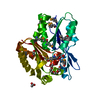
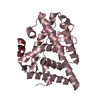
| Title | Structure of the Neurospora SET domain protein DIM-5, a histone lysine methyltransferase | |||||||||
 Components Components | Histone H3 methyltransferase DIM-5 | |||||||||
 Keywords Keywords | TRANSFERASE / DIM-5 / AdoMet-dependent Methyltransferase Histone H3 Lysine-9 Methylation | |||||||||
| Function / homology |  Function and homology information Function and homology information[histone H3]-lysine9 N-trimethyltransferase / histone H3K9 trimethyltransferase activity / histone methyltransferase activity / chromosome / double-stranded DNA binding / methylation / zinc ion binding / nucleus Similarity search - Function | |||||||||
| Biological species |  Neurospora crassa (fungus) Neurospora crassa (fungus) | |||||||||
| Method |  X-RAY DIFFRACTION / SYNCHROTRON / MAD / Resolution: 1.98 Å X-RAY DIFFRACTION / SYNCHROTRON / MAD / Resolution: 1.98 Å | |||||||||
 Authors Authors | Zhang, X. / Tamaru, H. / Khan, S.I. / Horton, J.R. / Keefe, L.J. / Selker, E.U. / Cheng, X. | |||||||||
 Citation Citation | Journal: Cell(Cambridge,Mass.) / Year: 2002 Title: Structure of the Neurospora SET domain protein DIM-5, a histone H3 lysine methyltransferase Authors: Zhang, X. / Tamaru, H. / Khan, S.I. / Horton, J.R. / Keefe, L.J. / Selker, E.U. / Cheng, X. | |||||||||
| History |
|
- Structure visualization
| Structure viewer | Molecule: MolmilJmol/JSmol |
|---|
- Downloads & links
-Download
| PDBx/mmCIF format | 1ml9.cif.gz | 66.4 KB | Display | PDBx/mmCIF format |
|---|---|---|---|---|
| PDB format | pdb1ml9.ent.gz | 46.8 KB | Display | PDB format |
| PDBx/mmJSON format | 1ml9.json.gz | Tree view | PDBx/mmJSON format | |
| Others |  Other downloads Other downloads |
-Validation report
| Arichive directory | https://data.pdbj.org/pub/pdb/validation_reports/ml/1ml9ftp://data.pdbj.org/pub/pdb/validation_reports/ml/1ml9 | HTTPS FTP |
|---|
-Related structure data
| Similar structure data |
|---|


-Links
 PDBj
PDBj

- Assembly
| Deposited unit | 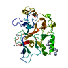
| ||||||||
|---|---|---|---|---|---|---|---|---|---|
| 1 |
| ||||||||
| Unit cell |
|
-Components
| #1: Protein | Mass: 34111.551 Da / Num. of mol.: 1 / Fragment: Residues 17-318 Source method: isolated from a genetically manipulated source Source: (gene. exp.) Neurospora crassa (fungus) / Plasmid: pXC379 / Species (production host): Escherichia coli / Production host:  References: GenBank: 17063801, UniProt: Q8X225*PLUS, histone-lysine N-methyltransferase | ||||||
|---|---|---|---|---|---|---|---|
| #2: Chemical |   Mass: 65.409 Da / Num. of mol.: 3 / Source method: obtained synthetically / Formula: Zn Mass: 65.409 Da / Num. of mol.: 3 / Source method: obtained synthetically / Formula: Zn#3: Chemical | ChemComp-UNL / Mass: 103.120 Da / Num. of mol.: 6 / Source method: obtained synthetically #4: Water | ChemComp-HOH / |  Mass: 18.015 Da / Num. of mol.: 111 / Source method: isolated from a natural source / Formula: H2O Mass: 18.015 Da / Num. of mol.: 111 / Source method: isolated from a natural source / Formula: H2OHas protein modification | N | |
-Experimental details
-Experiment
| Experiment | Method: X-RAY DIFFRACTION / Number of used crystals: 1 |
|---|
- Sample preparation
| Crystal | Density Matthews: 2.22 Å3/Da / Density % sol: 44.65 % | |||||||||||||||||||||||||||||||||||||||||||||||||||||||||||||||
|---|---|---|---|---|---|---|---|---|---|---|---|---|---|---|---|---|---|---|---|---|---|---|---|---|---|---|---|---|---|---|---|---|---|---|---|---|---|---|---|---|---|---|---|---|---|---|---|---|---|---|---|---|---|---|---|---|---|---|---|---|---|---|---|---|
| Crystal grow | Temperature: 289 K / Method: vapor diffusion, hanging drop / pH: 5.5 Details: ammonium sulfate, sodium citrate, pH 5.5, VAPOR DIFFUSION, HANGING DROP, temperature 289K | |||||||||||||||||||||||||||||||||||||||||||||||||||||||||||||||
| Crystal grow | *PLUS Temperature: 16 ℃ / pH: 9.8 | |||||||||||||||||||||||||||||||||||||||||||||||||||||||||||||||
| Components of the solutions | *PLUS
|
-Data collection
| Diffraction | Mean temperature: 100 K | ||||||||||||
|---|---|---|---|---|---|---|---|---|---|---|---|---|---|
| Diffraction source | Source: SYNCHROTRON / Site: APS  / Beamline: 17-ID / Wavelength: 1.0332, 1.2834, 1.2830 / Beamline: 17-ID / Wavelength: 1.0332, 1.2834, 1.2830 | ||||||||||||
| Detector | Type: ADSC QUANTUM 210 / Detector: CCD / Date: Feb 10, 2002 | ||||||||||||
| Radiation | Protocol: MAD / Monochromatic (M) / Laue (L): M / Scattering type: x-ray | ||||||||||||
| Radiation wavelength |
| ||||||||||||
| Reflection | Resolution: 1.98→24.83 Å / Num. all: 20963 / Num. obs: 20963 / % possible obs: 97 % / Observed criterion σ(F): 0 / Observed criterion σ(I): -3 / Redundancy: 5.37 % / Biso Wilson estimate: 26.9 Å2 / Rmerge(I) obs: 0.055 / Rsym value: 0.055 / Net I/σ(I): 15.4 | ||||||||||||
| Reflection shell | Resolution: 1.98→2.02 Å / Rmerge(I) obs: 0.276 / Mean I/σ(I) obs: 6.1 / Num. unique all: 1011 / Rsym value: 0.276 / % possible all: 95.8 | ||||||||||||
| Reflection | *PLUS % possible obs: 97 % / Num. measured all: 112569 / Rmerge(I) obs: 0.055 | ||||||||||||
| Reflection shell | *PLUS % possible obs: 95.8 % / Rmerge(I) obs: 0.276 |
- Processing
| Software |
| |||||||||||||||||||||||||
|---|---|---|---|---|---|---|---|---|---|---|---|---|---|---|---|---|---|---|---|---|---|---|---|---|---|---|
| Refinement | Method to determine structure: MAD / Resolution: 1.98→24.83 Å / Isotropic thermal model: Isotropic / Cross valid method: THROUGHOUT / σ(F): 2
| |||||||||||||||||||||||||
| Displacement parameters | Biso mean: 32.3 Å2 | |||||||||||||||||||||||||
| Refine analyze | Luzzati coordinate error obs: 0.24 Å / Luzzati d res low obs: 24.83 Å / Luzzati sigma a obs: 0.23 Å | |||||||||||||||||||||||||
| Refinement step | Cycle: LAST / Resolution: 1.98→24.83 Å
| |||||||||||||||||||||||||
| Refine LS restraints |
| |||||||||||||||||||||||||
| LS refinement shell | Resolution: 1.98→2.1 Å / Rfactor Rfree error: 0.015
| |||||||||||||||||||||||||
| Refinement | *PLUS Rfactor Rfree: 0.258 / Rfactor Rwork: 0.205 | |||||||||||||||||||||||||
| Solvent computation | *PLUS | |||||||||||||||||||||||||
| Displacement parameters | *PLUS | |||||||||||||||||||||||||
| Refine LS restraints | *PLUS
| |||||||||||||||||||||||||
| LS refinement shell | *PLUS Rfactor Rfree: 0.281 / Rfactor Rwork: 0.266 |
