 Movie
Movie Controller

[English] 日本語
 Yorodumi
- PDB-1mjl: METHIONINE REPRESSOR MUTANT (Q44K) COMPLEX WITH THE COREPRESSOR S... -
+ Open data

- Basic information
| Entry | Database: PDB / ID: 1mjl | ||||||
|---|---|---|---|---|---|---|---|
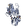
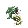
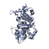
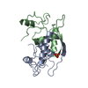
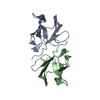
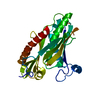
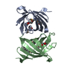
| Title | METHIONINE REPRESSOR MUTANT (Q44K) COMPLEX WITH THE COREPRESSOR SAM (S-ADENOSYL METHIONINE) FROM ESCHERICHIA COLI | ||||||
 Components Components | METHIONINE REPRESSOR PROTEIN METJ | ||||||
 Keywords Keywords | TRANSCRIPTION REGULATION / METJ / REPRESSOR / SHEET-HELIX-HELIX / SAM / S-ADENOSYL METHIONINE | ||||||
| Function / homology |  Function and homology information Function and homology information: / DNA-binding transcription factor activity / negative regulation of DNA-templated transcription / DNA binding / cytosol Similarity search - Function | ||||||
| Biological species |  | ||||||
| Method |  X-RAY DIFFRACTION / DIFFERENCE FOURIER METHODS / Resolution: 2.1 Å X-RAY DIFFRACTION / DIFFERENCE FOURIER METHODS / Resolution: 2.1 Å | ||||||
 Authors Authors | Garvie, C.W. / Phillips, S.E.V. | ||||||
 Citation Citation | Journal: Thesis, University of Leeds / Year: 1997 Title: Crystallographic studies of the methionine repressor-operator complex and the oc31 42kDa repressor Authors: Garvie, C.W. | ||||||
| History |
|
- Structure visualization
| Structure viewer | Molecule: MolmilJmol/JSmol |
|---|
- Downloads & links
-Download
| PDBx/mmCIF format | 1mjl.cif.gz | 57.1 KB | Display | PDBx/mmCIF format |
|---|---|---|---|---|
| PDB format | pdb1mjl.ent.gz | 41.5 KB | Display | PDB format |
| PDBx/mmJSON format | 1mjl.json.gz | Tree view | PDBx/mmJSON format | |
| Others |  Other downloads Other downloads |
-Validation report
| Arichive directory | https://data.pdbj.org/pub/pdb/validation_reports/mj/1mjlftp://data.pdbj.org/pub/pdb/validation_reports/mj/1mjl | HTTPS FTP |
|---|
-Related structure data
| Related structure data |  1cmcS S: Starting model for refinement |
|---|---|
| Similar structure data |


-Links
 PDBj
PDBj
- Assembly
| Deposited unit | 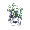
| ||||||||
|---|---|---|---|---|---|---|---|---|---|
| 1 |
| ||||||||
| Unit cell |
|
-Components
| #1: Protein | Mass: 12028.607 Da / Num. of mol.: 2 / Mutation: Q44K Source method: isolated from a genetically manipulated source Source: (gene. exp.) #2: Chemical |   Mass: 398.437 Da / Num. of mol.: 2 / Source method: obtained synthetically / Formula: C15H22N6O5S Mass: 398.437 Da / Num. of mol.: 2 / Source method: obtained synthetically / Formula: C15H22N6O5S#3: Water | ChemComp-HOH / |  Mass: 18.015 Da / Num. of mol.: 112 / Source method: isolated from a natural source / Formula: H2O Mass: 18.015 Da / Num. of mol.: 112 / Source method: isolated from a natural source / Formula: H2O |
|---|
-Experimental details
-Experiment
| Experiment | Method: X-RAY DIFFRACTION / Number of used crystals: 1 |
|---|
- Sample preparation
| Crystal | Density Matthews: 1.95 Å3/Da / Density % sol: 37 % |
|---|---|
| Crystal grow | pH: 7 Details: PROTEIN (10MG/ML) + SAM (1MG/ ML) WAS CRYSTALLIZED FROM 10-30% PEG 600, 100MM SODIUM CACODYLATE BUFFER, PH 4.6-5.2., pH 7.0 PH range: 4.6-5.2 |
| Crystal grow | *PLUS Method: unknown |
-Data collection
| Diffraction | Mean temperature: 293 K |
|---|---|
| Diffraction source | Source: ROTATING ANODE / Type: RIGAKU RUH2R / Wavelength: 1.5418 |
| Detector | Type: SIEMENS / Detector: AREA DETECTOR / Date: Mar 1, 1995 |
| Radiation | Monochromator: GRAPHITE(002) / Monochromatic (M) / Laue (L): M / Scattering type: x-ray |
| Radiation wavelength | Wavelength: 1.5418 Å / Relative weight: 1 |
| Reflection | Resolution: 2.1→28 Å / Num. obs: 10414 / % possible obs: 97.9 % / Observed criterion σ(I): 3 / Redundancy: 2.9 % / Biso Wilson estimate: 13.9 Å2 / Rmerge(I) obs: 0.049 / Net I/σ(I): 10.4 |
| Reflection shell | Resolution: 2.1→2.21 Å / Redundancy: 2.4 % / Rmerge(I) obs: 0.049 / Mean I/σ(I) obs: 2.6 / Rsym value: 0.19 / % possible all: 86.9 |
- Processing
| Software |
| ||||||||||||||||||||||||||||||||||||||||||||||||||||||||||||
|---|---|---|---|---|---|---|---|---|---|---|---|---|---|---|---|---|---|---|---|---|---|---|---|---|---|---|---|---|---|---|---|---|---|---|---|---|---|---|---|---|---|---|---|---|---|---|---|---|---|---|---|---|---|---|---|---|---|---|---|---|---|
| Refinement | Method to determine structure: DIFFERENCE FOURIER METHODS Starting model: PDB ENTRY 1CMC Resolution: 2.1→28 Å / Rfactor Rfree error: 0.006 / Data cutoff high absF: 10000000 / Data cutoff low absF: 0.001 / Isotropic thermal model: RESTRAINED / Cross valid method: THROUGHOUT / σ(F): 2 / Details: BULK SOLVENT MODEL USED
| ||||||||||||||||||||||||||||||||||||||||||||||||||||||||||||
| Displacement parameters | Biso mean: 27.6 Å2 | ||||||||||||||||||||||||||||||||||||||||||||||||||||||||||||
| Refine analyze |
| ||||||||||||||||||||||||||||||||||||||||||||||||||||||||||||
| Refinement step | Cycle: LAST / Resolution: 2.1→28 Å
| ||||||||||||||||||||||||||||||||||||||||||||||||||||||||||||
| Refine LS restraints |
| ||||||||||||||||||||||||||||||||||||||||||||||||||||||||||||
| LS refinement shell | Resolution: 2.1→2.18 Å / Rfactor Rfree error: 0.03 / Total num. of bins used: 10
| ||||||||||||||||||||||||||||||||||||||||||||||||||||||||||||
| Xplor file |
| ||||||||||||||||||||||||||||||||||||||||||||||||||||||||||||
| Software | *PLUS Version: 3.86 / Classification: refinement | ||||||||||||||||||||||||||||||||||||||||||||||||||||||||||||
| Refinement | *PLUS Rfactor Rfree: 0.197 / Rfactor Rwork: 0.193 | ||||||||||||||||||||||||||||||||||||||||||||||||||||||||||||
| Solvent computation | *PLUS | ||||||||||||||||||||||||||||||||||||||||||||||||||||||||||||
| Displacement parameters | *PLUS | ||||||||||||||||||||||||||||||||||||||||||||||||||||||||||||
| Refine LS restraints | *PLUS
| ||||||||||||||||||||||||||||||||||||||||||||||||||||||||||||
| LS refinement shell | *PLUS Rfactor Rfree: 0.287 / Rfactor Rwork: 0.241 |
