 Movie
Movie Controller

[English] 日本語
 Yorodumi
- PDB-1cmc: THREE DIMENSIONAL CRYSTAL STRUCTURES OF E. COLI MET REPRESSOR WIT... -
+ Open data

- Basic information
| Entry | Database: PDB / ID: 1cmc | ||||||
|---|---|---|---|---|---|---|---|
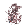
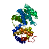
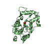
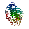
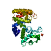
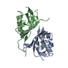
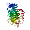
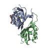
| Title | THREE DIMENSIONAL CRYSTAL STRUCTURES OF E. COLI MET REPRESSOR WITH AND WITHOUT COREPRESSOR | ||||||
 Components Components | MET REPRESSOR | ||||||
 Keywords Keywords | DNA-BINDING REGULATORY PROTEIN | ||||||
| Function / homology |  Function and homology information Function and homology information: / DNA-binding transcription factor activity / negative regulation of DNA-templated transcription / DNA binding / cytosol Similarity search - Function | ||||||
| Biological species |  | ||||||
| Method |  X-RAY DIFFRACTION / Resolution: 1.8 Å X-RAY DIFFRACTION / Resolution: 1.8 Å | ||||||
 Authors Authors | Somers, W.S. / Phillips, S.E.V. | ||||||
 Citation Citation | Journal: Nature / Year: 1989 Title: Three-dimensional crystal structures of Escherichia coli met repressor with and without corepressor. Authors: Rafferty, J.B. / Somers, W.S. / Saint-Girons, I. / Phillips, S.E. | ||||||
| History |
|
- Structure visualization
| Structure viewer | Molecule: MolmilJmol/JSmol |
|---|
- Downloads & links
-Download
| PDBx/mmCIF format | 1cmc.cif.gz | 61.3 KB | Display | PDBx/mmCIF format |
|---|---|---|---|---|
| PDB format | pdb1cmc.ent.gz | 45.1 KB | Display | PDB format |
| PDBx/mmJSON format | 1cmc.json.gz | Tree view | PDBx/mmJSON format | |
| Others |  Other downloads Other downloads |
-Validation report
| Arichive directory | https://data.pdbj.org/pub/pdb/validation_reports/cm/1cmcftp://data.pdbj.org/pub/pdb/validation_reports/cm/1cmc | HTTPS FTP |
|---|
-Related structure data


-Links
 PDBj
PDBj
- Assembly
| Deposited unit | 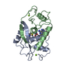
| ||||||||
|---|---|---|---|---|---|---|---|---|---|
| 1 |
| ||||||||
| Unit cell |
| ||||||||
| Atom site foot note | 1: THE IDENTITY OF RESIDUES 106 A AND 106 B IS UNKNOWN. EACH INTERACTS WITH HIS 14, GLU 19, A SYMMETRY RELATED GLU 103, AND THE C-TERMINUS. THE MAGNITUDE OF THE DENSITY PEAK SUGGESTS ATOMIC NUMBER IN ...1: THE IDENTITY OF RESIDUES 106 A AND 106 B IS UNKNOWN. EACH INTERACTS WITH HIS 14, GLU 19, A SYMMETRY RELATED GLU 103, AND THE C-TERMINUS. THE MAGNITUDE OF THE DENSITY PEAK SUGGESTS ATOMIC NUMBER IN RANGE 25 - 35. IT IS POSSIBLY ARSENIC FROM BUFFER. IN REFINEMENT IT WAS MODELLED BY A MAGNESIUM ION WITH OCCUPANCY 2.0. |
-Components
| #1: Protein | Mass: 12027.557 Da / Num. of mol.: 2 Source method: isolated from a genetically manipulated source Source: (gene. exp.) #2: Chemical |   Mass: 24.305 Da / Num. of mol.: 2 / Source method: obtained synthetically / Formula: Mg Mass: 24.305 Da / Num. of mol.: 2 / Source method: obtained synthetically / Formula: Mg#3: Chemical |   Mass: 398.437 Da / Num. of mol.: 2 / Source method: obtained synthetically / Formula: C15H22N6O5S Mass: 398.437 Da / Num. of mol.: 2 / Source method: obtained synthetically / Formula: C15H22N6O5S#4: Water | ChemComp-HOH / |  Mass: 18.015 Da / Num. of mol.: 206 / Source method: isolated from a natural source / Formula: H2O Mass: 18.015 Da / Num. of mol.: 206 / Source method: isolated from a natural source / Formula: H2ONonpolymer details | THE IDENTITY OF RESIDUES 106 A AND 106 B IS UNKNOWN. EACH INTERACTS WITH HIS 14, GLU 19, A SYMMETRY ...THE IDENTITY OF RESIDUES 106 A AND 106 B IS UNKNOWN. EACH INTERACTS WITH HIS 14, GLU 19, A SYMMETRY RELATED GLU 103, AND THE C-TERMINUS. THE MAGNITUDE OF THE DENSITY PEAK SUGGESTS ATOMIC NUMBER IN RANGE 25 - 35. IT IS POSSIBLY ARSENIC FROM BUFFER. IN REFINEMENT | |
|---|
-Experimental details
-Experiment
| Experiment | Method: X-RAY DIFFRACTION |
|---|
- Sample preparation
| Crystal | Density Matthews: 2.76 Å3/Da / Density % sol: 55.51 % | ||||||||||||||||||||||||
|---|---|---|---|---|---|---|---|---|---|---|---|---|---|---|---|---|---|---|---|---|---|---|---|---|---|
| Crystal grow | *PLUS Method: microdialysis / pH: 6.2 | ||||||||||||||||||||||||
| Components of the solutions | *PLUS
|
-Data collection
| Radiation | Scattering type: x-ray |
|---|---|
| Radiation wavelength | Relative weight: 1 |
- Processing
| Software | Name: PROLSQ / Classification: refinement | |||||||||||||||||||||||||||||||||||||||||||||||||||||||||||||||
|---|---|---|---|---|---|---|---|---|---|---|---|---|---|---|---|---|---|---|---|---|---|---|---|---|---|---|---|---|---|---|---|---|---|---|---|---|---|---|---|---|---|---|---|---|---|---|---|---|---|---|---|---|---|---|---|---|---|---|---|---|---|---|---|---|
| Refinement | Rfactor obs: 0.183 / Highest resolution: 1.8 Å | |||||||||||||||||||||||||||||||||||||||||||||||||||||||||||||||
| Refinement step | Cycle: LAST / Highest resolution: 1.8 Å
| |||||||||||||||||||||||||||||||||||||||||||||||||||||||||||||||
| Refine LS restraints |
| |||||||||||||||||||||||||||||||||||||||||||||||||||||||||||||||
| Software | *PLUS Name: PROLSQ / Classification: refinement | |||||||||||||||||||||||||||||||||||||||||||||||||||||||||||||||
| Refinement | *PLUS Highest resolution: 1.8 Å / σ(F): 3 / Rfactor obs: 0.183 | |||||||||||||||||||||||||||||||||||||||||||||||||||||||||||||||
| Solvent computation | *PLUS | |||||||||||||||||||||||||||||||||||||||||||||||||||||||||||||||
| Displacement parameters | *PLUS Biso mean: 6.2 Å2 | |||||||||||||||||||||||||||||||||||||||||||||||||||||||||||||||
| Refine LS restraints | *PLUS
|
