 Movie
Movie Controller

[English] 日本語
 Yorodumi
- PDB-1mi3: 1.8 Angstrom structure of xylose reductase from Candida tenuis in... -
+ Open data

- Basic information
| Entry | Database: PDB / ID: 1mi3 | ||||||
|---|---|---|---|---|---|---|---|
| Title | 1.8 Angstrom structure of xylose reductase from Candida tenuis in complex with NAD | ||||||
 Components Components | xylose reductase | ||||||
 Keywords Keywords | OXIDOREDUCTASE / aldo-keto reductase / beta-alpha barrel / dimer | ||||||
| Function / homology |  Function and homology information Function and homology informationD-xylose reductase [NAD(P)H] / D-xylose reductase (NADPH) activity / D-xylose catabolic process Similarity search - Function | ||||||
| Biological species |  Candida tenuis (fungus) Candida tenuis (fungus) | ||||||
| Method |  X-RAY DIFFRACTION / SYNCHROTRON / MOLECULAR REPLACEMENT / Resolution: 1.8 Å X-RAY DIFFRACTION / SYNCHROTRON / MOLECULAR REPLACEMENT / Resolution: 1.8 Å | ||||||
 Authors Authors | Kavanagh, K.L. / Klimacek, M. / Nidetzky, B. / Wilson, D.K. | ||||||
 Citation Citation | Journal: Biochem.J. / Year: 2003 Title: Structure of xylose reductase bound to NAD+ and the basis for single and dual co-substrate specificity in family 2 aldo-keto reductases Authors: Kavanagh, K.L. / Klimacek, M. / Nidetzky, B. / Wilson, D.K. | ||||||
| History |
|
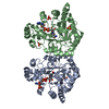
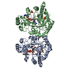
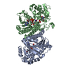
- Structure visualization
| Structure viewer | Molecule: MolmilJmol/JSmol |
|---|
- Downloads & links
-Download
| PDBx/mmCIF format | 1mi3.cif.gz | 277.9 KB | Display | PDBx/mmCIF format |
|---|---|---|---|---|
| PDB format | pdb1mi3.ent.gz | 224.6 KB | Display | PDB format |
| PDBx/mmJSON format | 1mi3.json.gz | Tree view | PDBx/mmJSON format | |
| Others |  Other downloads Other downloads |
-Validation report
| Summary document | 1mi3_validation.pdf.gz | 1.4 MB | Display | wwPDB validaton report |
|---|---|---|---|---|
| Full document | 1mi3_full_validation.pdf.gz | 1.5 MB | Display | |
| Data in XML | 1mi3_validation.xml.gz | 55.3 KB | Display | |
| Data in CIF | 1mi3_validation.cif.gz | 79.6 KB | Display | |
| Arichive directory | https://data.pdbj.org/pub/pdb/validation_reports/mi/1mi3ftp://data.pdbj.org/pub/pdb/validation_reports/mi/1mi3 | HTTPS FTP |
-Related structure data
| Related structure data | |
|---|---|
| Similar structure data |


-Links
 PDBj
PDBj
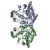
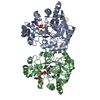
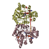
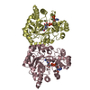
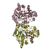
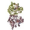
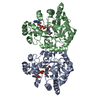
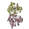
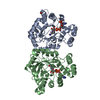
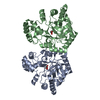
- Assembly
| Deposited unit | 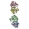
| ||||||||||||||||||
|---|---|---|---|---|---|---|---|---|---|---|---|---|---|---|---|---|---|---|---|
| 1 | 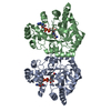
| ||||||||||||||||||
| 2 | 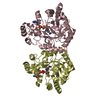
| ||||||||||||||||||
| 3 | 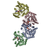
| ||||||||||||||||||
| Unit cell |
| ||||||||||||||||||
| Components on special symmetry positions |
|
-Components
| #1: Protein | Mass: 36062.199 Da / Num. of mol.: 4 Source method: isolated from a genetically manipulated source Source: (gene. exp.) Candida tenuis (fungus) / Gene: xylR / Plasmid: pBEAct.1i / Species (production host): Escherichia coli / Production host:  #2: Chemical | ChemComp-NAD /   Mass: 663.425 Da / Num. of mol.: 4 / Source method: obtained synthetically / Formula: C21H27N7O14P2 / Comment: NAD*YM Mass: 663.425 Da / Num. of mol.: 4 / Source method: obtained synthetically / Formula: C21H27N7O14P2 / Comment: NAD*YM#3: Water | ChemComp-HOH / |  Mass: 18.015 Da / Num. of mol.: 873 / Source method: isolated from a natural source / Formula: H2O Mass: 18.015 Da / Num. of mol.: 873 / Source method: isolated from a natural source / Formula: H2O |
|---|
-Experimental details
-Experiment
| Experiment | Method: X-RAY DIFFRACTION / Number of used crystals: 1 |
|---|
- Sample preparation
| Crystal | Density Matthews: 2.95 Å3/Da / Density % sol: 58 % | ||||||||||||||||||||||||||||||||||||||||||||||||||||||
|---|---|---|---|---|---|---|---|---|---|---|---|---|---|---|---|---|---|---|---|---|---|---|---|---|---|---|---|---|---|---|---|---|---|---|---|---|---|---|---|---|---|---|---|---|---|---|---|---|---|---|---|---|---|---|---|
| Crystal grow | Temperature: 293 K / Method: vapor diffusion, hanging drop / pH: 5.6 Details: 32% PEG-ME 5K, 350 mM ammonium sulfate, 100 mM sodium citrate, pH 5.6, VAPOR DIFFUSION, HANGING DROP, temperature 293K | ||||||||||||||||||||||||||||||||||||||||||||||||||||||
| Crystal grow | *PLUS Method: vapor diffusion | ||||||||||||||||||||||||||||||||||||||||||||||||||||||
| Components of the solutions | *PLUS
|
-Data collection
| Diffraction | Mean temperature: 100 K |
|---|---|
| Diffraction source | Source: SYNCHROTRON / Site: SSRL  / Beamline: BL9-1 / Wavelength: 0.97 Å / Beamline: BL9-1 / Wavelength: 0.97 Å |
| Detector | Type: MARRESEARCH / Detector: IMAGE PLATE / Date: Jan 2, 2002 |
| Radiation | Protocol: SINGLE WAVELENGTH / Monochromatic (M) / Laue (L): M / Scattering type: x-ray |
| Radiation wavelength | Wavelength: 0.97 Å / Relative weight: 1 |
| Reflection | Resolution: 1.8→30 Å / Num. all: 164527 / Num. obs: 162718 / % possible obs: 98.9 % / Observed criterion σ(I): 2 / Rmerge(I) obs: 0.063 / Net I/σ(I): 13 |
| Reflection shell | Resolution: 1.8→1.83 Å / Rmerge(I) obs: 0.359 / Mean I/σ(I) obs: 3 / % possible all: 98.2 |
| Reflection | *PLUS Highest resolution: 1.8 Å / Num. obs: 162716 / Num. measured all: 728917 |
| Reflection shell | *PLUS % possible obs: 98.2 % / Mean I/σ(I) obs: 3.02 |
- Processing
| Software |
| |||||||||||||||||||||||||
|---|---|---|---|---|---|---|---|---|---|---|---|---|---|---|---|---|---|---|---|---|---|---|---|---|---|---|
| Refinement | Method to determine structure: MOLECULAR REPLACEMENT Starting model: unpublished model of XR mutant Resolution: 1.8→30 Å / Cross valid method: THROUGHOUT / σ(F): 0 / Stereochemistry target values: Engh & Huber
| |||||||||||||||||||||||||
| Displacement parameters |
| |||||||||||||||||||||||||
| Refinement step | Cycle: LAST / Resolution: 1.8→30 Å
| |||||||||||||||||||||||||
| Refine LS restraints |
| |||||||||||||||||||||||||
| Refinement | *PLUS Highest resolution: 1.8 Å / Lowest resolution: 30 Å / Num. reflection obs: 154541 / Rfactor Rfree: 0.212 / Rfactor Rwork: 0.183 | |||||||||||||||||||||||||
| Solvent computation | *PLUS | |||||||||||||||||||||||||
| Displacement parameters | *PLUS | |||||||||||||||||||||||||
| Refine LS restraints | *PLUS Type: c_angle_deg / Dev ideal: 1.7 |
