 Movie
Movie Controller

[English] 日本語
 Yorodumi
- PDB-1mhm: Crystal structure of S-adenosylmethionine decarboxylase from potato -
+ Open data

- Basic information
| Entry | Database: PDB / ID: 1mhm | |||||||||
|---|---|---|---|---|---|---|---|---|---|---|
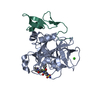
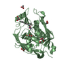
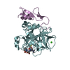
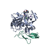
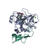
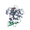
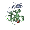
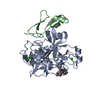
| Title | Crystal structure of S-adenosylmethionine decarboxylase from potato | |||||||||
 Components Components | (S-adenosylmethionine decarboxylase) x 2 | |||||||||
 Keywords Keywords | LYASE / covalent pyruvoyl group | |||||||||
| Function / homology |  Function and homology information Function and homology informationplant organ development / adenosylmethionine decarboxylase / adenosylmethionine decarboxylase activity / spermine biosynthetic process / spermidine biosynthetic process / cytosol Similarity search - Function | |||||||||
| Biological species |  | |||||||||
| Method |  X-RAY DIFFRACTION / MOLECULAR REPLACEMENT / Resolution: 2.3 Å X-RAY DIFFRACTION / MOLECULAR REPLACEMENT / Resolution: 2.3 Å | |||||||||
 Authors Authors | Bennett, E.M. / Ekstrom, J.L. / Pegg, A.E. / Ealick, S.E. | |||||||||
 Citation Citation | Journal: Biochemistry / Year: 2002 Title: Monomeric S-Adenosylmethionine Decarboxylase from Plants Provides an Alternative to Putrescine Stimulation Authors: Bennett, E.M. / Ekstrom, J.L. / Pegg, A.E. / Ealick, S.E. | |||||||||
| History |
|
- Structure visualization
| Structure viewer | Molecule: MolmilJmol/JSmol |
|---|
- Downloads & links
-Download
| PDBx/mmCIF format | 1mhm.cif.gz | 74.5 KB | Display | PDBx/mmCIF format |
|---|---|---|---|---|
| PDB format | pdb1mhm.ent.gz | 54.2 KB | Display | PDB format |
| PDBx/mmJSON format | 1mhm.json.gz | Tree view | PDBx/mmJSON format | |
| Others |  Other downloads Other downloads |
-Validation report
| Arichive directory | https://data.pdbj.org/pub/pdb/validation_reports/mh/1mhmftp://data.pdbj.org/pub/pdb/validation_reports/mh/1mhm | HTTPS FTP |
|---|
-Related structure data
| Related structure data | |
|---|---|
| Similar structure data |


-Links
 PDBj
PDBj- Assembly
| Deposited unit | 
| ||||||||
|---|---|---|---|---|---|---|---|---|---|
| 1 |
| ||||||||
| Unit cell |
|
-Components
| #1: Protein | Mass: 7886.826 Da / Num. of mol.: 1 / Fragment: beta chain Source method: isolated from a genetically manipulated source Source: (gene. exp.)  References: UniProt: Q04694, adenosylmethionine decarboxylase |
|---|---|
| #2: Protein | Mass: 31882.322 Da / Num. of mol.: 1 / Fragment: alpha chain Source method: isolated from a genetically manipulated source Source: (gene. exp.) References: UniProt: Q04694, adenosylmethionine decarboxylase |
| #3: Water | ChemComp-HOH /  Mass: 18.015 Da / Num. of mol.: 84 / Source method: isolated from a natural source / Formula: H2O Mass: 18.015 Da / Num. of mol.: 84 / Source method: isolated from a natural source / Formula: H2O |
| Has protein modification | Y |
-Experimental details
-Experiment
| Experiment | Method: X-RAY DIFFRACTION / Number of used crystals: 1 |
|---|
- Sample preparation
| Crystal | Density Matthews: 2.14 Å3/Da / Density % sol: 42.48 % | ||||||||||||||||||||||||
|---|---|---|---|---|---|---|---|---|---|---|---|---|---|---|---|---|---|---|---|---|---|---|---|---|---|
| Crystal grow | Temperature: 290 K / Method: vapor diffusion, hanging drop / pH: 5.6 Details: trisodium citrate, isopropanol, PEG 4000, pH 5.6, VAPOR DIFFUSION, HANGING DROP, temperature 290K | ||||||||||||||||||||||||
| Crystal grow | *PLUS Temperature: 17 ℃ | ||||||||||||||||||||||||
| Components of the solutions | *PLUS
|
-Data collection
| Diffraction | Mean temperature: 77 K |
|---|---|
| Diffraction source | Source: ROTATING ANODE / Type: RIGAKU / Wavelength: 1.54 Å |
| Detector | Type: MARRESEARCH / Detector: IMAGE PLATE / Date: Jan 1, 2000 |
| Radiation | Protocol: SINGLE WAVELENGTH / Monochromatic (M) / Laue (L): M / Scattering type: x-ray |
| Radiation wavelength | Wavelength: 1.54 Å / Relative weight: 1 |
| Reflection | Resolution: 2.1→99 Å / Num. all: 20754 / Num. obs: 18907 / % possible obs: 91.1 % / Observed criterion σ(F): 0 / Observed criterion σ(I): 0 |
| Reflection shell | Resolution: 2.1→2.18 Å / Rsym value: 0.409 / % possible all: 79.1 |
| Reflection | *PLUS Num. obs: 17049 / % possible obs: 90.1 % / Redundancy: 3.8 % / Rmerge(I) obs: 0.087 |
| Reflection shell | *PLUS Highest resolution: 2.26 Å / Lowest resolution: 2.37 Å / % possible obs: 69.6 % / Redundancy: 2.8 % / Num. unique obs: 1084 / Rmerge(I) obs: 0.318 / Mean I/σ(I) obs: 2.9 |
- Processing
| Software |
| |||||||||||||||||||||
|---|---|---|---|---|---|---|---|---|---|---|---|---|---|---|---|---|---|---|---|---|---|---|
| Refinement | Method to determine structure: MOLECULAR REPLACEMENT / Resolution: 2.3→50 Å / σ(F): 0 / σ(I): 0 / Stereochemistry target values: Engh & Huber
| |||||||||||||||||||||
| Refine analyze | Luzzati coordinate error free: 0.4 Å | |||||||||||||||||||||
| Refinement step | Cycle: LAST / Resolution: 2.3→50 Å
| |||||||||||||||||||||
| Refine LS restraints |
| |||||||||||||||||||||
| LS refinement shell | Resolution: 2.3→2.38 Å
| |||||||||||||||||||||
| Xplor file |
| |||||||||||||||||||||
| Refinement | *PLUS Lowest resolution: 500 Å | |||||||||||||||||||||
| Solvent computation | *PLUS | |||||||||||||||||||||
| Displacement parameters | *PLUS | |||||||||||||||||||||
| Refine LS restraints | *PLUS
| |||||||||||||||||||||
| LS refinement shell | *PLUS Highest resolution: 2.3 Å / Rfactor Rfree: 0.398 / Num. reflection Rfree: 61 / Num. reflection obs: 1070 |
