 Movie
Movie Controller

[English] 日本語
 Yorodumi
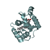
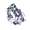
- PDB-1mg4: STRUCTURE OF N-TERMINAL DOUBLECORTIN DOMAIN FROM DCLK: WILD TYPE ... -
+ Open data

- Basic information
| Entry | Database: PDB / ID: 1mg4 | ||||||
|---|---|---|---|---|---|---|---|
| Title | STRUCTURE OF N-TERMINAL DOUBLECORTIN DOMAIN FROM DCLK: WILD TYPE PROTEIN | ||||||
 Components Components | DOUBLECORTIN-LIKE KINASE (N-TERMINAL DOMAIN) | ||||||
 Keywords Keywords | TRANSFERASE / DCX Domain / Doublecortin-like Kinase / Microtubule Bundling / Cortex Development | ||||||
| Function / homology |  Function and homology information Function and homology informationaxon extension / endosomal transport / protein localization to nucleus / central nervous system development / response to virus / nervous system development / protein phosphorylation / protein kinase activity / non-specific serine/threonine protein kinase / intracellular signal transduction ...axon extension / endosomal transport / protein localization to nucleus / central nervous system development / response to virus / nervous system development / protein phosphorylation / protein kinase activity / non-specific serine/threonine protein kinase / intracellular signal transduction / protein serine kinase activity / protein serine/threonine kinase activity / ATP binding / plasma membrane / cytoplasm Similarity search - Function | ||||||
| Biological species |  Homo sapiens (human) Homo sapiens (human) | ||||||
| Method |  X-RAY DIFFRACTION / SYNCHROTRON / MOLECULAR REPLACEMENT / Resolution: 1.504 Å X-RAY DIFFRACTION / SYNCHROTRON / MOLECULAR REPLACEMENT / Resolution: 1.504 Å | ||||||
 Authors Authors | Kim, M.H. / Cierpickil, T. / Derewenda, U. / Krowarsch, D. / Feng, Y. / Devedjiev, Y. / Dauter, Z. / Walsh, C.A. / Otlewski, J. / Bushweller, J.H. / Derewenda, Z. | ||||||
 Citation Citation | Journal: Nat.Struct.Biol. / Year: 2003 Title: The DCX-domain Tandems of Doublecortin and Doublecortin-like Kinase Authors: Kim, M.H. / Cierpickil, T. / Derewenda, U. / Krowarsch, D. / Feng, Y. / Devedjiev, Y. / Dauter, Z. / Walsh, C.A. / Otlewski, J. / Bushweller, J.H. / Derewenda, Z. | ||||||
| History |
|
- Structure visualization
| Structure viewer | Molecule: MolmilJmol/JSmol |
|---|
- Downloads & links
-Download
| PDBx/mmCIF format | 1mg4.cif.gz | 59.4 KB | Display | PDBx/mmCIF format |
|---|---|---|---|---|
| PDB format | pdb1mg4.ent.gz | 43.4 KB | Display | PDB format |
| PDBx/mmJSON format | 1mg4.json.gz | Tree view | PDBx/mmJSON format | |
| Others |  Other downloads Other downloads |
-Validation report
| Arichive directory | https://data.pdbj.org/pub/pdb/validation_reports/mg/1mg4ftp://data.pdbj.org/pub/pdb/validation_reports/mg/1mg4 | HTTPS FTP |
|---|
-Related structure data
| Related structure data |  1mfwSC 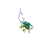 1mjdC S: Starting model for refinement C: citing same article ( |
|---|---|
| Similar structure data |


-Links
 PDBj
PDBj- Assembly
| Deposited unit | 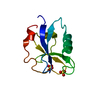
| ||||||||
|---|---|---|---|---|---|---|---|---|---|
| 1 |
| ||||||||
| Unit cell |
| ||||||||
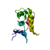
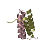
| Details | BIOLOGICAL ASSEMBLY IS A TANDEM OF N-TERMINAL AND C-TERMINAL DOMAINS |
-Components
| #1: Protein | Mass: 12877.623 Da / Num. of mol.: 1 / Fragment: (N-Terminal Domain), Residues 49-154 Source method: isolated from a genetically manipulated source Source: (gene. exp.) Homo sapiens (human) / Production host:  | ||
|---|---|---|---|
| #2: Chemical |   Mass: 96.063 Da / Num. of mol.: 2 / Source method: obtained synthetically / Formula: SO4 Mass: 96.063 Da / Num. of mol.: 2 / Source method: obtained synthetically / Formula: SO4#3: Water | ChemComp-HOH / |  Mass: 18.015 Da / Num. of mol.: 140 / Source method: isolated from a natural source / Formula: H2O Mass: 18.015 Da / Num. of mol.: 140 / Source method: isolated from a natural source / Formula: H2O |
-Experimental details
-Experiment
| Experiment | Method: X-RAY DIFFRACTION / Number of used crystals: 1 |
|---|
- Sample preparation
| Crystal | Density Matthews: 1.95 Å3/Da / Density % sol: 36.4 % | ||||||||||||||||||||||||
|---|---|---|---|---|---|---|---|---|---|---|---|---|---|---|---|---|---|---|---|---|---|---|---|---|---|
| Crystal grow | Temperature: 294 K / Method: vapor diffusion, sitting drop / pH: 5 Details: CITRATE BUFFER, AMMONIUM SULFATE, pH 5.0, VAPOR DIFFUSION, SITTING DROP, temperature 294K | ||||||||||||||||||||||||
| Crystal grow | *PLUS Temperature: 21 ℃ | ||||||||||||||||||||||||
| Components of the solutions | *PLUS
|
-Data collection
| Diffraction | Mean temperature: 100 K |
|---|---|
| Diffraction source | Source: SYNCHROTRON / Site: APS  / Beamline: 19-ID / Wavelength: 0.9187 Å / Beamline: 19-ID / Wavelength: 0.9187 Å |
| Detector | Type: ADSC QUANTUM 4 / Detector: CCD / Date: Mar 8, 2002 / Details: Double crystal focusing mirrors |
| Radiation | Monochromator: Si (III) / Protocol: SINGLE WAVELENGTH / Monochromatic (M) / Laue (L): M / Scattering type: x-ray |
| Radiation wavelength | Wavelength: 0.9187 Å / Relative weight: 1 |
| Reflection | Resolution: 1.5→50 Å / Num. all: 15460 / Num. obs: 15460 / % possible obs: 96.8 % / Observed criterion σ(I): -3 / Redundancy: 3.1 % / Biso Wilson estimate: 13.7 Å2 / Rmerge(I) obs: 0.084 / Net I/σ(I): 14.1 |
| Reflection shell | Resolution: 1.5→1.55 Å / Redundancy: 2.1 % / Rmerge(I) obs: 0.286 / Mean I/σ(I) obs: 2.5 / Num. unique all: 1368 / % possible all: 85.7 |
| Reflection | *PLUS Highest resolution: 1.5 Å / Lowest resolution: 50 Å / Num. measured all: 47701 |
| Reflection shell | *PLUS Highest resolution: 1.5 Å / Lowest resolution: 1.55 Å / % possible obs: 85.7 % |
- Processing
| Software |
| ||||||||||||||||||||||||||||||||||||||||||||||||||||||||||||||||||||||||||||||||||||||||||||||||||||||||||||||||||||||||
|---|---|---|---|---|---|---|---|---|---|---|---|---|---|---|---|---|---|---|---|---|---|---|---|---|---|---|---|---|---|---|---|---|---|---|---|---|---|---|---|---|---|---|---|---|---|---|---|---|---|---|---|---|---|---|---|---|---|---|---|---|---|---|---|---|---|---|---|---|---|---|---|---|---|---|---|---|---|---|---|---|---|---|---|---|---|---|---|---|---|---|---|---|---|---|---|---|---|---|---|---|---|---|---|---|---|---|---|---|---|---|---|---|---|---|---|---|---|---|---|---|---|
| Refinement | Method to determine structure: MOLECULAR REPLACEMENT Starting model: PDB ENTRY 1MFW Resolution: 1.504→19.28 Å / Cor.coef. Fo:Fc: 0.969 / Cor.coef. Fo:Fc free: 0.956 / SU B: 3.745 / SU ML: 0.071 / Cross valid method: THROUGHOUT / σ(F): 1 / ESU R: 0.095 / ESU R Free: 0.074 / Stereochemistry target values: MAXIMUM LIKELIHOOD / Details: HYDROGENS HAVE BEEN ADDED IN THE RIDING POSITIONS
| ||||||||||||||||||||||||||||||||||||||||||||||||||||||||||||||||||||||||||||||||||||||||||||||||||||||||||||||||||||||||
| Solvent computation | Ion probe radii: 0.8 Å / Shrinkage radii: 0.8 Å / VDW probe radii: 1.4 Å / Solvent model: BABINET MODEL WITH MASK | ||||||||||||||||||||||||||||||||||||||||||||||||||||||||||||||||||||||||||||||||||||||||||||||||||||||||||||||||||||||||
| Displacement parameters | Biso mean: 15.754 Å2
| ||||||||||||||||||||||||||||||||||||||||||||||||||||||||||||||||||||||||||||||||||||||||||||||||||||||||||||||||||||||||
| Refinement step | Cycle: LAST / Resolution: 1.504→19.28 Å
| ||||||||||||||||||||||||||||||||||||||||||||||||||||||||||||||||||||||||||||||||||||||||||||||||||||||||||||||||||||||||
| Refine LS restraints |
| ||||||||||||||||||||||||||||||||||||||||||||||||||||||||||||||||||||||||||||||||||||||||||||||||||||||||||||||||||||||||
| LS refinement shell | Resolution: 1.504→1.543 Å / Total num. of bins used: 20 /
| ||||||||||||||||||||||||||||||||||||||||||||||||||||||||||||||||||||||||||||||||||||||||||||||||||||||||||||||||||||||||
| Refinement | *PLUS Highest resolution: 1.5 Å / Lowest resolution: 20 Å / Rfactor Rfree: 0.187 / Rfactor Rwork: 0.153 | ||||||||||||||||||||||||||||||||||||||||||||||||||||||||||||||||||||||||||||||||||||||||||||||||||||||||||||||||||||||||
| Solvent computation | *PLUS | ||||||||||||||||||||||||||||||||||||||||||||||||||||||||||||||||||||||||||||||||||||||||||||||||||||||||||||||||||||||||
| Displacement parameters | *PLUS | ||||||||||||||||||||||||||||||||||||||||||||||||||||||||||||||||||||||||||||||||||||||||||||||||||||||||||||||||||||||||
| Refine LS restraints | *PLUS
|
