 Movie
Movie Controller


+ Open data

- Basic information
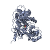
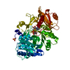
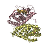
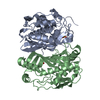
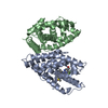
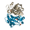
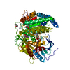
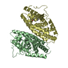
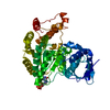
| Entry | Database: PDB / ID: 1mdf | ||||||
|---|---|---|---|---|---|---|---|
| Title | CRYSTAL STRUCTURE OF DhbE IN ABSENCE OF SUBSTRATE | ||||||
 Components Components | 2,3-dihydroxybenzoate-AMP ligase | ||||||
 Keywords Keywords | LIGASE / adenylation domain / peptide synthetase / antibiotic biosynthesis / siderophore formation | ||||||
| Function / homology |  Function and homology information Function and homology information2,3-dihydroxybenzoate-[aryl-carrier protein] ligase / 2,3-dihydroxybenzoate--[aryl-carrier protein] ligase activity / siderophore biosynthetic process / ATP binding / cytoplasm Similarity search - Function | ||||||
| Biological species |  | ||||||
| Method |  X-RAY DIFFRACTION / MOLECULAR REPLACEMENT / Resolution: 2.5 Å X-RAY DIFFRACTION / MOLECULAR REPLACEMENT / Resolution: 2.5 Å | ||||||
 Authors Authors | May, J.J. / Kessler, N. / Marahiel, M.A. / Stubbs, M.T. | ||||||
 Citation Citation | Journal: Proc.Natl.Acad.Sci.USA / Year: 2002 Title: Crystal structure of DhbE, an archetype for aryl acid activating domains of modular nonribosomal peptide synthetases. Authors: May, J.J. / Kessler, N. / Marahiel, M.A. / Stubbs, M.T. | ||||||
| History |
| ||||||
| Remark 999 | SEQUENCE Author states the sequence has recently been deposited at NCBI with the acquisition number ...SEQUENCE Author states the sequence has recently been deposited at NCBI with the acquisition number bankit484943. |
- Structure visualization
| Structure viewer | Molecule: MolmilJmol/JSmol |
|---|
- Downloads & links
-Download
| PDBx/mmCIF format | 1mdf.cif.gz | 115.3 KB | Display | PDBx/mmCIF format |
|---|---|---|---|---|
| PDB format | pdb1mdf.ent.gz | 89.9 KB | Display | PDB format |
| PDBx/mmJSON format | 1mdf.json.gz | Tree view | PDBx/mmJSON format | |
| Others |  Other downloads Other downloads |
-Validation report
| Arichive directory | https://data.pdbj.org/pub/pdb/validation_reports/md/1mdfftp://data.pdbj.org/pub/pdb/validation_reports/md/1mdf | HTTPS FTP |
|---|
-Related structure data
| Related structure data | 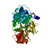 1md9SC  1mdbC S: Starting model for refinement C: citing same article ( |
|---|---|
| Similar structure data |


-Links
 PDBj
PDBj
- Assembly
| Deposited unit | 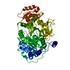
| ||||||||
|---|---|---|---|---|---|---|---|---|---|
| 1 |
| ||||||||
| Unit cell |
|
-Components
| #1: Protein | Mass: 59988.137 Da / Num. of mol.: 1 Source method: isolated from a genetically manipulated source Source: (gene. exp.) References: UniProt: P40871, Ligases; Forming carbon-nitrogen bonds; Acid-amino-acid ligases (peptide synthases) | ||
|---|---|---|---|
| #2: Chemical |   Mass: 96.063 Da / Num. of mol.: 2 / Source method: obtained synthetically / Formula: SO4 Mass: 96.063 Da / Num. of mol.: 2 / Source method: obtained synthetically / Formula: SO4#3: Water | ChemComp-HOH / |  Mass: 18.015 Da / Num. of mol.: 93 / Source method: isolated from a natural source / Formula: H2O Mass: 18.015 Da / Num. of mol.: 93 / Source method: isolated from a natural source / Formula: H2O |
-Experimental details
-Experiment
| Experiment | Method: X-RAY DIFFRACTION |
|---|
- Sample preparation
| Crystal | Density Matthews: 2.2 Å3/Da / Density % sol: 44.06 % | |||||||||||||||||||||||||||||||||||
|---|---|---|---|---|---|---|---|---|---|---|---|---|---|---|---|---|---|---|---|---|---|---|---|---|---|---|---|---|---|---|---|---|---|---|---|---|
| Crystal grow | *PLUS Temperature: 18 ℃ / pH: 8.6 / Method: vapor diffusion, sitting drop | |||||||||||||||||||||||||||||||||||
| Components of the solutions | *PLUS
|
-Data collection
| Detector | Date: Mar 10, 2001 |
|---|---|
| Radiation | Protocol: SINGLE WAVELENGTH / Monochromatic (M) / Laue (L): M / Scattering type: x-ray |
| Radiation wavelength | Relative weight: 1 |
| Reflection | Resolution: 2.5→100 Å / Num. obs: 16523 / Observed criterion σ(I): 0 |
| Reflection | *PLUS Highest resolution: 2.5 Å / Num. obs: 17099 / % possible obs: 96.5 % / Num. measured all: 42630 / Rmerge(I) obs: 0.088 |
| Reflection shell | *PLUS % possible obs: 92.4 % / Rmerge(I) obs: 0.318 / Mean I/σ(I) obs: 2.68 |
- Processing
| Software |
| ||||||||||||||||
|---|---|---|---|---|---|---|---|---|---|---|---|---|---|---|---|---|---|
| Refinement | Method to determine structure: MOLECULAR REPLACEMENT Starting model: PDB ENTRY 1MD9 Resolution: 2.5→100 Å / Cross valid method: THROUGHOUT / σ(F): 0
| ||||||||||||||||
| Refinement step | Cycle: LAST / Resolution: 2.5→100 Å
| ||||||||||||||||
| Refinement | *PLUS Rfactor Rfree: 0.275 / Rfactor Rwork: 0.204 / % reflection Rfree: 10 % | ||||||||||||||||
| Solvent computation | *PLUS | ||||||||||||||||
| Displacement parameters | *PLUS | ||||||||||||||||
| Refine LS restraints | *PLUS
|
